Copyright
©2011 Baishideng Publishing Group Co.
World J Gastrointest Surg. Mar 27, 2011; 3(3): 39-42
Published online Mar 27, 2011. doi: 10.4240/wjgs.v3.i3.39
Published online Mar 27, 2011. doi: 10.4240/wjgs.v3.i3.39
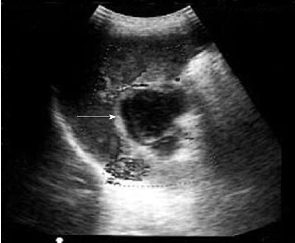
Figure 1 Ultrasound oblique scan through the long axis of the portal vein.
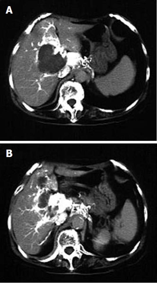
Figure 2 Abdominal computed tomography scan with contrast.
A: A hypovascular mass in the porta hepatis; B: Developed collateral vessels around the mass.
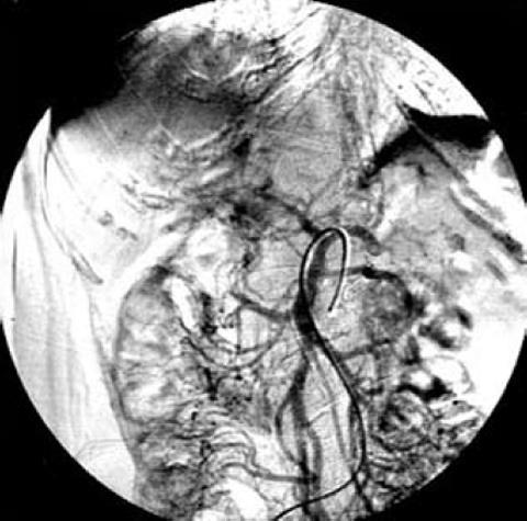
Figure 3 Portal venogram (digital subtraction angiogram) after selective superior mesenteric arteriography.
- Citation: Ishimura K, Otani T, Wakabayashi H, Okano K, Goda F, Suzuki Y. A case report of extrahepatic portal vein aneurysm with thrombosis. World J Gastrointest Surg 2011; 3(3): 39-42
- URL: https://www.wjgnet.com/1948-9366/full/v3/i3/39.htm
- DOI: https://dx.doi.org/10.4240/wjgs.v3.i3.39


