Copyright
©2011 Baishideng Publishing Group Co.
World J Gastrointest Surg. Dec 27, 2011; 3(12): 201-203
Published online Dec 27, 2011. doi: 10.4240/wjgs.v3.i12.201
Published online Dec 27, 2011. doi: 10.4240/wjgs.v3.i12.201
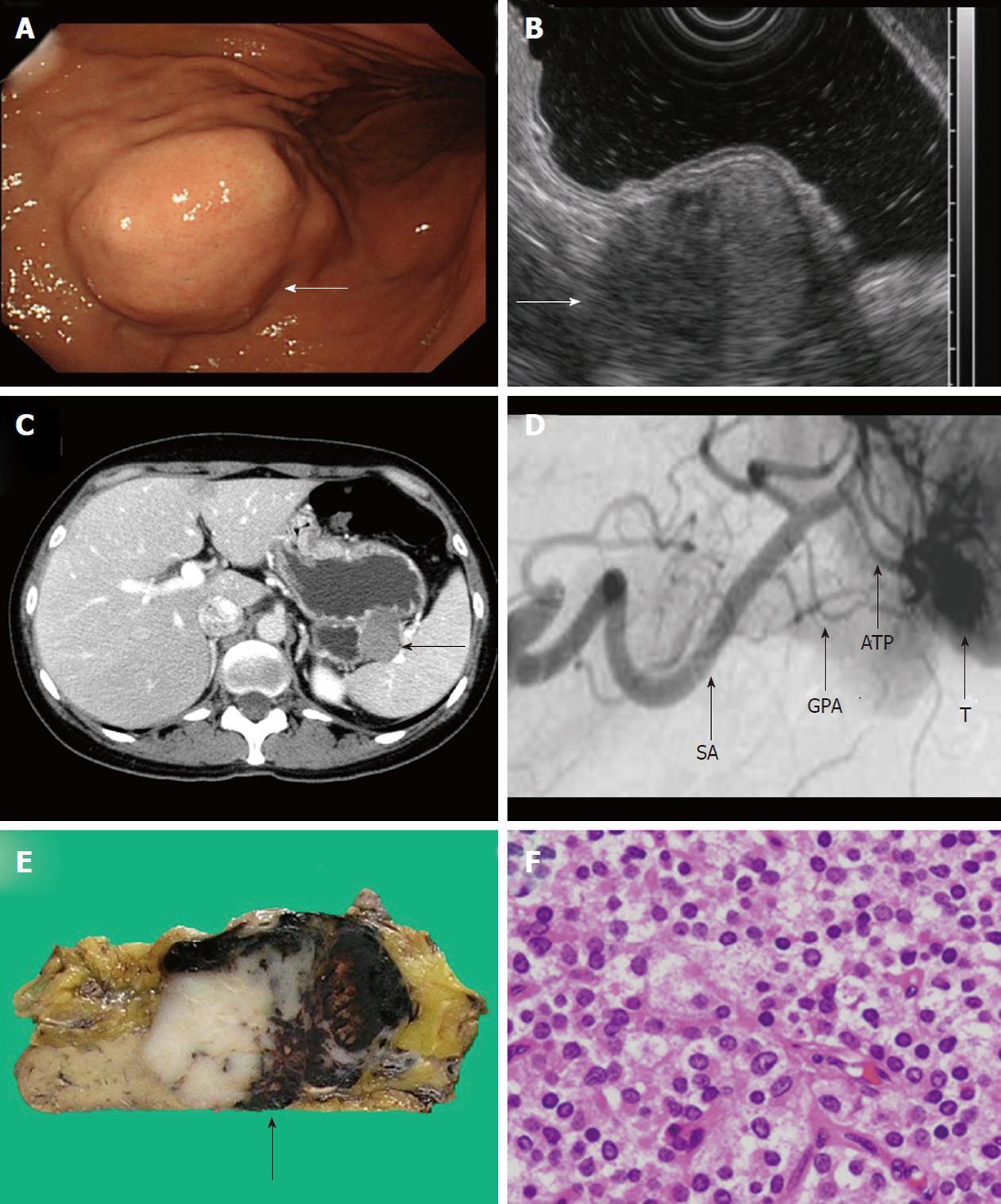
Figure 1 Figures of the patient.
A: Preoperative imaging Elevated tumor (arrow) without mucosal abnormality was shown on endoscopy; B: Endoscopic ultrasonography revealed that the tumor had heterogeneous internal echoes and continuity of the muscularis propria; C: Abdominal CT showed the tumor close to splenic hilum; D: Angiography showed tumor vessels originated from pancreatic arteries; E: The tumor was solid with focal necrosis and hemorrhage; F: Histological examination revealed tumor cells with round to oval nuclei. GPA: Greater pancreatic artery; ATP: Artery to tail of the pancreas; SA: Splenic artery; T: Tumor.
- Citation: Furuhashi S, Takamori H, Abe S, Nakahara O, Tanaka H, Horino K, Beppu T, Iyama KI, Baba H. Solid-pseudopapillary pancreatic tumor, mimicking submucosal tumor of the stomach: A case report. World J Gastrointest Surg 2011; 3(12): 201-203
- URL: https://www.wjgnet.com/1948-9366/full/v3/i12/201.htm
- DOI: https://dx.doi.org/10.4240/wjgs.v3.i12.201


