Copyright
©2010 Baishideng Publishing Group Co.
World J Gastrointest Surg. Sep 27, 2010; 2(9): 291-294
Published online Sep 27, 2010. doi: 10.4240/wjgs.v2.i9.291
Published online Sep 27, 2010. doi: 10.4240/wjgs.v2.i9.291
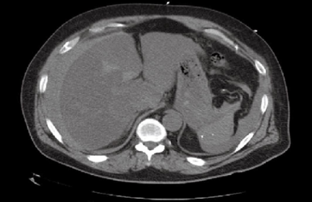
Figure 1 Computed tomography showing acute peritoneal hemorrhage.
Fluid is prominent surrounding the second and third portions of the duodenum and pancreatic head, and perihepatic regions.
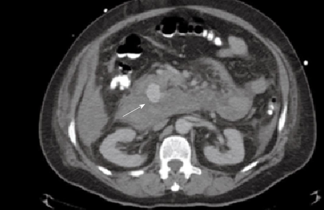
Figure 2 Abdominal contrast enhanced computed tomography showing retroperitoneal aneurysm which is suspected to be arising from the gastroduodenal artery or one of its branches (arrow).
Aneurysm measures 3.1 cm × 2.5 cm.
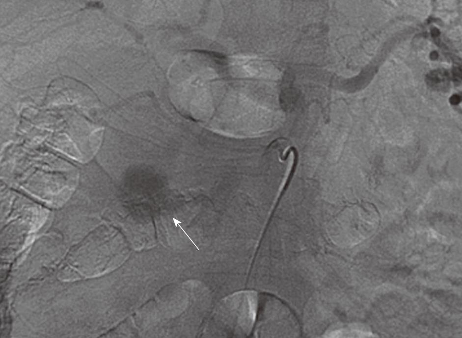
Figure 3 Abdominal angiography, selective superior mesenteric artery angiography showing the gastroduodenal artery aneurysm (arrow).
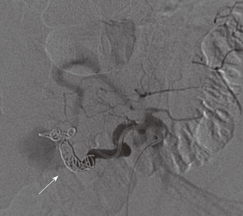
Figure 4 Post embolization angiography (arrow) showing no residual filling of the gastroduodenal artery aneurysm.
- Citation: Harris K, Chalhoub M, Koirala A. Gastroduodenal artery aneurysm rupture in hospitalized patients: An overlooked diagnosis. World J Gastrointest Surg 2010; 2(9): 291-294
- URL: https://www.wjgnet.com/1948-9366/full/v2/i9/291.htm
- DOI: https://dx.doi.org/10.4240/wjgs.v2.i9.291


