Copyright
©2010 Baishideng.
World J Gastrointest Surg. Jul 27, 2010; 2(7): 251-254
Published online Jul 27, 2010. doi: 10.4240/wjgs.v2.i7.251
Published online Jul 27, 2010. doi: 10.4240/wjgs.v2.i7.251
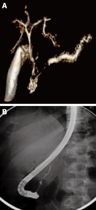
Figure 1 A magnetic resonant (A) and endoscopic retrograde cholangio-pancreatography (B) showing complete ductal disruption.
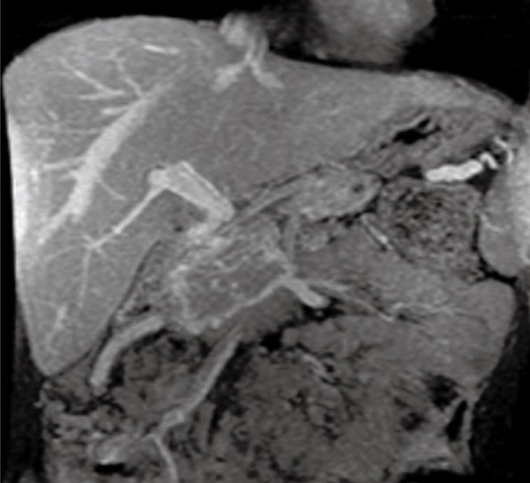
Figure 2 Magnetic resonance imaging film showing portosplenic thrombosis, gastric varices and pancreatoduodenal collaterals.
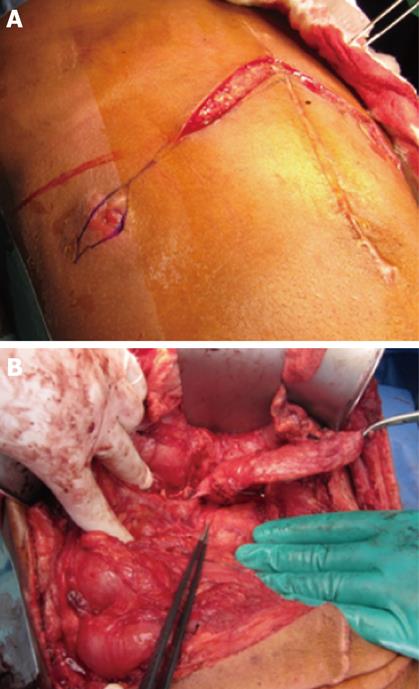
Figure 3 Cutaneous orifice of the pancreatic fistula is circled (A) and the operative view of the inner orifice at the injured neck of pancreas (B).
- Citation: Bojal SA, Leung KF, Meshikhes AWN. Traumatic pancreatic fistula with sinistral portal hypertension: Surgical management. World J Gastrointest Surg 2010; 2(7): 251-254
- URL: https://www.wjgnet.com/1948-9366/full/v2/i7/251.htm
- DOI: https://dx.doi.org/10.4240/wjgs.v2.i7.251


