Copyright
©2010 Baishideng.
World J Gastrointest Surg. May 27, 2010; 2(5): 157-164
Published online May 27, 2010. doi: 10.4240/wjgs.v2.i5.157
Published online May 27, 2010. doi: 10.4240/wjgs.v2.i5.157
Figure 1 Animal model.
A: Right side. *Right Iliac Vessels; B: Left side. *Ureter; C: Left side. *Kidney, upper pole; **Adrenal gland; ***Tail of the Pancreas; D: Left side. *Adrenal gland.
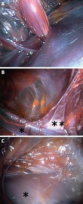
Figure 2 Cadaver model.
A: Left side. Pudendal nerve entering the Alcock canal; B: Left side. *External Iliac Vein; **Inferior Epigastric Vein; C: Left side. *Kidney, lower pole.
- Citation: Allemann P, Perretta S, Asakuma M, Dallemagne B, Marescaux J. NOTES new frontier: Natural orifice approach to retroperitoneal disease. World J Gastrointest Surg 2010; 2(5): 157-164
- URL: https://www.wjgnet.com/1948-9366/full/v2/i5/157.htm
- DOI: https://dx.doi.org/10.4240/wjgs.v2.i5.157


