Copyright
©2010 Baishideng.
World J Gastrointest Surg. Apr 27, 2010; 2(4): 137-142
Published online Apr 27, 2010. doi: 10.4240/wjgs.v2.i4.137
Published online Apr 27, 2010. doi: 10.4240/wjgs.v2.i4.137
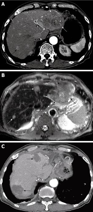
Figure 1 Successful radiofrequency ablation (RFA) combined with hepatic resection for multifocal hepatocellular carcinomas (HCCs) in a 74-year-old man.
A: Contrast-enhanced transverse helical CT scan obtained during the arterial phase before treatment shows two HCCs in the both hepatic lobes. There are 11.0-cm-diameter HCC (white arrows) in left lateral segment and 1.1-cm-diameter HCC (black arrow) in liver segment 7; B: Ferucarbotran-enhanced T2-weighted fast spin echo MR image shows another 1.5-cm-diameter HCC (long white arrow) in liver segment 4. He underwent resection for a large tumor and concurrent RFA for two small tumors; C. Contrast-enhanced transverse helical CT scan obtained 1 mo after combined hepatectomy and RFA shows two round ablation zones (arrows) of low attenuation that suggests technical success of ablation. At 4 mo after treatment, multiple metastatic tumors were found in the both lungs (not shown).
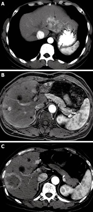
Figure 2 Successful RFA of intrahepatic recurrent HCC in a 49-year-old man.
A: Contrast-enhanced transverse helical CT scan obtained during the arterial phase before hepatectomy shows a 5.7-cm-diameter HCC (arrows) in left lateral segment; B: At 5 years after left lateral segmentectomy, Gd-EOB-DTPA-enhanced T1-weighted MR image obtained during the arterial phase before RFA shows a 1.0-cm-diameter recurrent HCC (arrow) in liver segment 8; C: Contrast-enhanced transverse helical CT scan obtained 1 h after RFA shows a round ablation zone (arrows) of low attenuation with peripheral rim hyperemia. The patient has been alive without recurrence for 14 mo.
- Citation: Choi D, Lim HK, Rhim H. Concurrent and subsequent radiofrequency ablation combined with hepatectomy for hepatocellular carcinomas. World J Gastrointest Surg 2010; 2(4): 137-142
- URL: https://www.wjgnet.com/1948-9366/full/v2/i4/137.htm
- DOI: https://dx.doi.org/10.4240/wjgs.v2.i4.137


