Copyright
©2010 Baishideng Publishing Group Co.
World J Gastrointest Surg. Dec 27, 2010; 2(12): 402-404
Published online Dec 27, 2010. doi: 10.4240/wjgs.v2.i12.402
Published online Dec 27, 2010. doi: 10.4240/wjgs.v2.i12.402
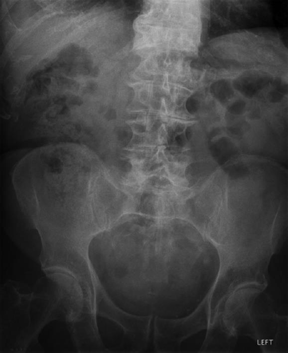
Figure 1 Abdominal radiograph appearances were non-specific.
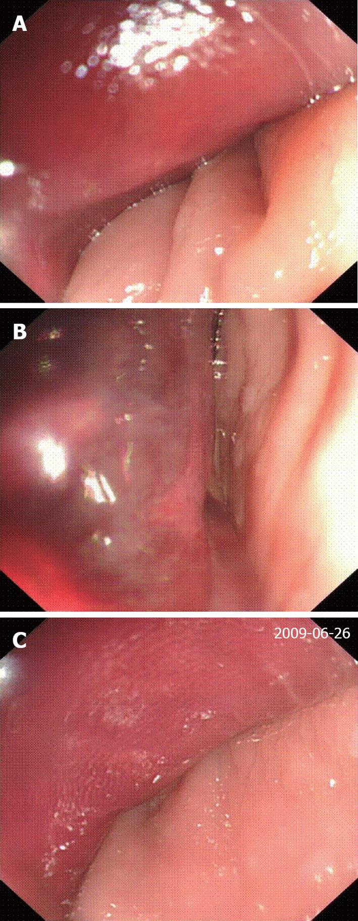
Figure 2 Images taken at flexible sigmoidoscopy demonstrating an extra-luminal mass putting pressure of the anterior rectal wall.
A: Compression of the anterior rectal wall; B: Haematoma due to gangrenous small bowel loops; C: Pressure within the pelvic cavity acting on the rectum.
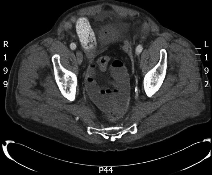
Figure 3 Computed tomography scan demonstrating the presence of dilated, fluid-filled loops of small bowel clumped together within the pelvis.
- Citation: Mownah OA, Hamady ZZ, Rogers MJ, Shah SG, Vani DH. Small bowel obstruction presenting with a rectal haematoma. World J Gastrointest Surg 2010; 2(12): 402-404
- URL: https://www.wjgnet.com/1948-9366/full/v2/i12/402.htm
- DOI: https://dx.doi.org/10.4240/wjgs.v2.i12.402


