Copyright
©2010 Baishideng Publishing Group Co.
World J Gastrointest Surg. Dec 27, 2010; 2(12): 395-401
Published online Dec 27, 2010. doi: 10.4240/wjgs.v2.i12.395
Published online Dec 27, 2010. doi: 10.4240/wjgs.v2.i12.395
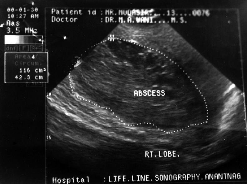
Figure 1 Ultrasonographic picture showing abscess in the right lobe of the liver.
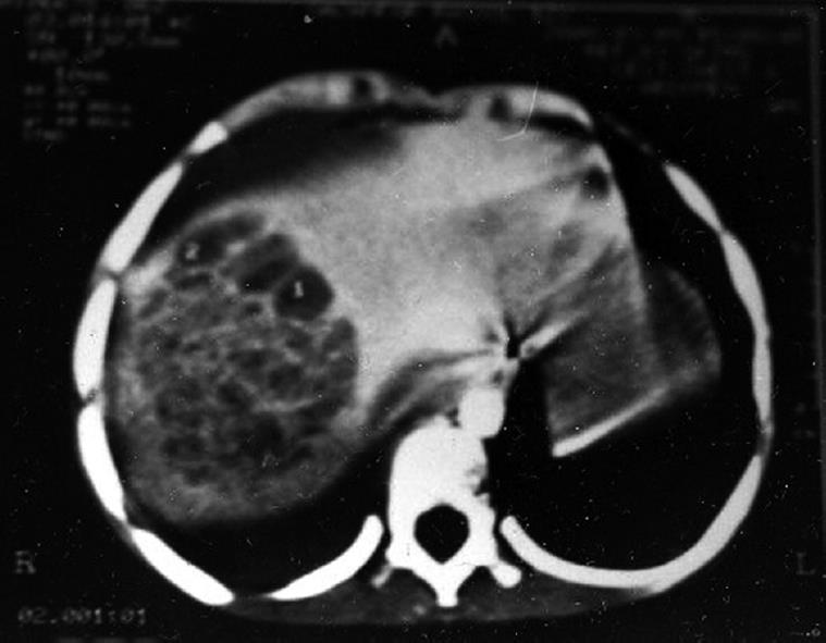
Figure 2 Computed tomography scan picture showing abscess in the right lobe of the liver.
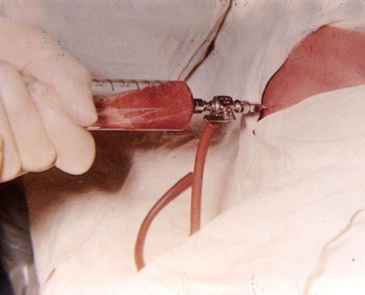
Figure 3 Percutaneous drainage of liver abscess being carried out.
- Citation: Malik AA, Bari SU, Rouf KA, Wani KA. Pyogenic liver abscess: Changing patterns in approach. World J Gastrointest Surg 2010; 2(12): 395-401
- URL: https://www.wjgnet.com/1948-9366/full/v2/i12/395.htm
- DOI: https://dx.doi.org/10.4240/wjgs.v2.i12.395


