Copyright
©2010 Baishideng Publishing Group Co.
World J Gastrointest Surg. Oct 27, 2010; 2(10): 319-323
Published online Oct 27, 2010. doi: 10.4240/wjgs.v2.i10.319
Published online Oct 27, 2010. doi: 10.4240/wjgs.v2.i10.319
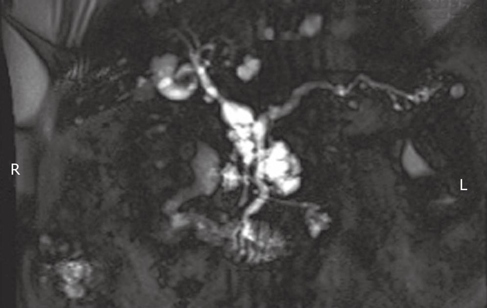
Figure 1 MRCP demonstrating intraductal papillary mucinous neoplasm in the head of the pancreas and uncinate, with dilated main duct > 6 mm, and multiple dilated side branches, likely representing mixed-type intraductal papillary mucinous neoplasm.
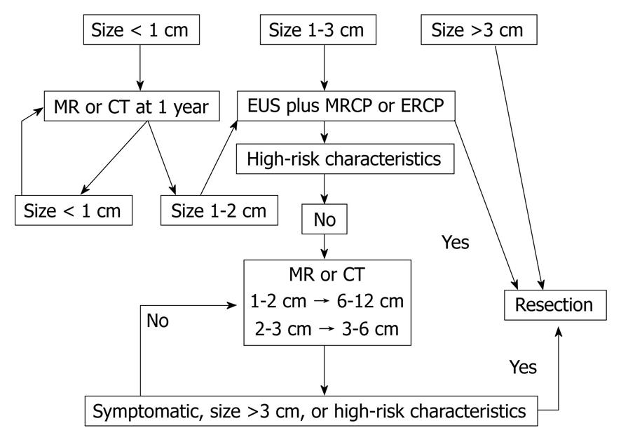
Figure 2 Management algorithm for intraductal papillary mucinous neoplasm (branch duct)[12].
CT: Computed tomography.
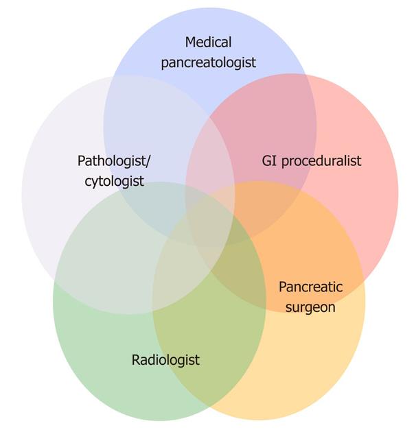
Figure 3 Multidisciplinary components in the management of pancreatic cystic neoplasms.
- Citation: Kent TS, Jr CMV, Callery MP. Intraductal papillary mucinous neoplasm and the pancreatic incidentaloma. World J Gastrointest Surg 2010; 2(10): 319-323
- URL: https://www.wjgnet.com/1948-9366/full/v2/i10/319.htm
- DOI: https://dx.doi.org/10.4240/wjgs.v2.i10.319


