Copyright
©The Author(s) 2025.
World J Gastrointest Surg. Aug 27, 2025; 17(8): 108656
Published online Aug 27, 2025. doi: 10.4240/wjgs.v17.i8.108656
Published online Aug 27, 2025. doi: 10.4240/wjgs.v17.i8.108656
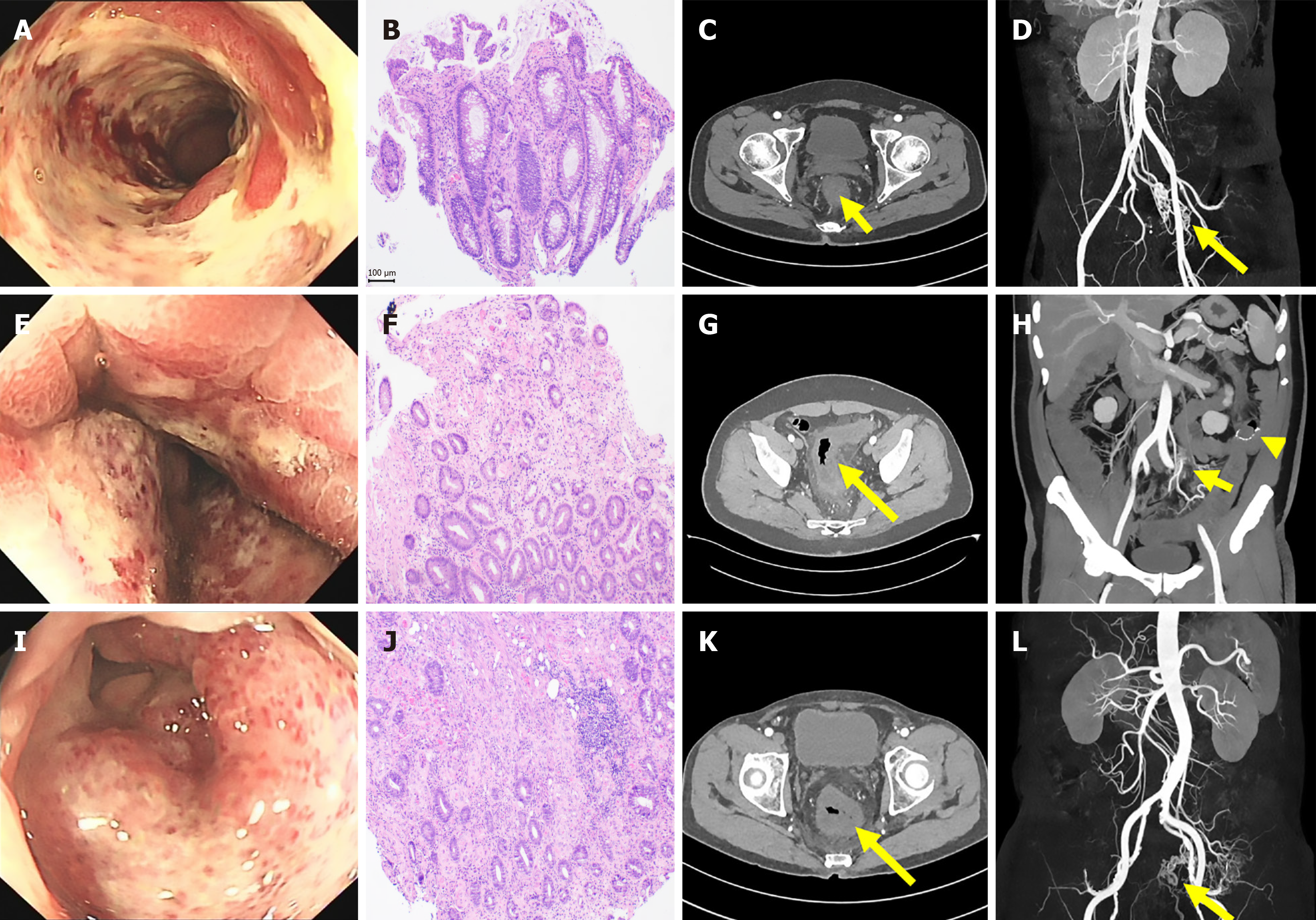
Figure 1 Endoscopies, pathology and imaging of the three cases.
A: Endoscopy from case 1 showed diffuse ulceration in the rectum; B: Pathology of the rectum showed epithelial damage and exfoliation, crypt distortion, stromal hemorrhage with local hyaline degeneration, capillary dilation in the stroma, and local thickening of the vascular wall; C: Contrast-enhanced abdominal computed tomography (CT) revealed circumferential wall thickening of the rectum; D: Abdominal CT angiography (CTA) demonstrated an arteriovenous fistula in the inferior mesenteric vessels; E: Endoscopic image of case 2 illustrated diffuse swelling of the rectosigmoid mucosa; F: Epithelial damage and exfoliation, crypt distortion, stromal hemorrhage with local hyaline degeneration, capillary dilation in the stroma, and local thickening of the vascular wall; G: Contrast-enhanced abdominal CT depicted edematous thickening of the rectosigmoid wall with surrounding exudates; H: Abdominal CTA revealed an arteriovenous fistula in the inferior mesenteric vessels; I: Endoscopic image of case 3 displayed diffuse mucosal roughness, congestion, and ulceration from the colonic anastomosis to the anal verge; J: Epithelial damage and exfoliation, crypt distortion, stromal hemorrhage with local hyaline degeneration, capillary dilation in the stroma, and local thickening of the vascular wall; K: Contrast-enhanced abdominal CT indicated swelling of the colorectal wall distal to the anastomosis with surrounding blurry exudation; L: Abdominal CTA showed an arteriovenous fistula in the inferior mesenteric vessels.
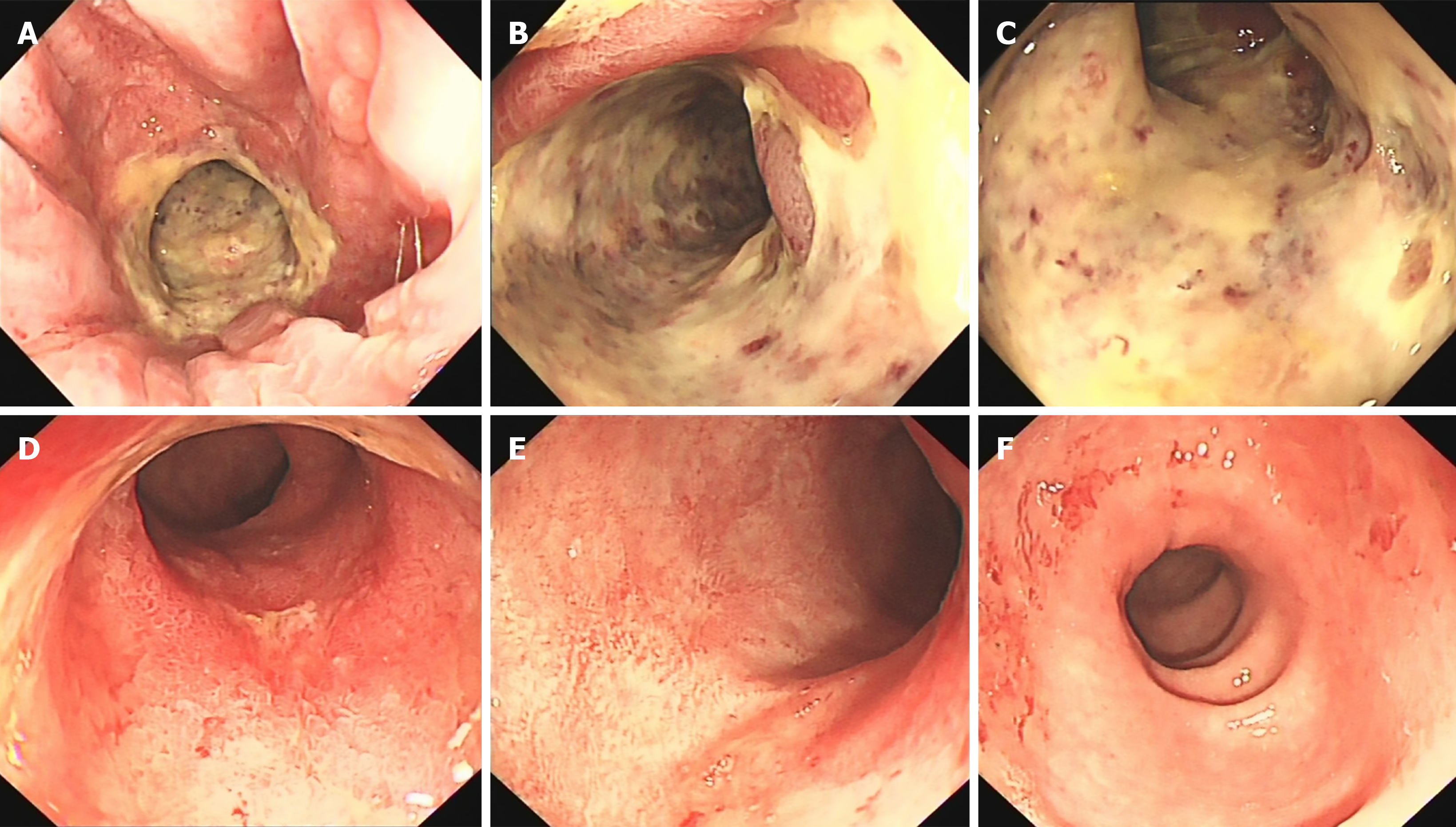
Figure 2 Pre-embolization and post-embolization endoscopy.
A-C: Pre-treatment endoscopic images of the patient show diffuse ulceration of the rectal mucosa; D-F: Post-embolization endoscopic review indicates significant improvement of the mucosal lesions.
Figure 3 Digital subtraction angiography imaging during inferior mesenteric artery angiography and embolization in case 2 and case 3.
A: Angiography from case 2, showing dilation of the inferior mesenteric artery, early visualization and significant dilation of the inferior mesenteric vein, along with a disordered vasculature; B: Post-embolization imaging from case 2, showing the disappearance of the local disordered vasculature and the visualization of the inferior mesenteric vein; C: Angiography from case 3, showing dilation of the inferior mesenteric artery, early visualization and significant dilation of the inferior mesenteric vein, along with a disordered vascular network; D: Post-embolization review from case 3, showing the disappearance of the local disordered vasculature and the visualization of the inferior mesenteric vein.
- Citation: Mei SB, Liu J, Wang Y, Hu P, Cao Q. Inferior mesenteric arteriovenous fistula: Three case reports. World J Gastrointest Surg 2025; 17(8): 108656
- URL: https://www.wjgnet.com/1948-9366/full/v17/i8/108656.htm
- DOI: https://dx.doi.org/10.4240/wjgs.v17.i8.108656


