Copyright
©The Author(s) 2025.
World J Gastrointest Surg. Aug 27, 2025; 17(8): 107558
Published online Aug 27, 2025. doi: 10.4240/wjgs.v17.i8.107558
Published online Aug 27, 2025. doi: 10.4240/wjgs.v17.i8.107558
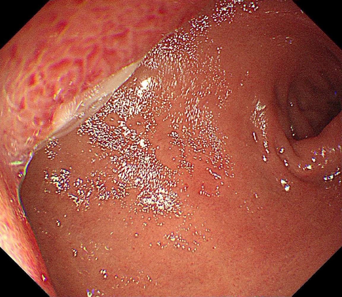
Figure 1
Gastroscopy showed a mucosal bulge in the duodenal bulb, involving the pylorus, and the surface of the lesion was congested.
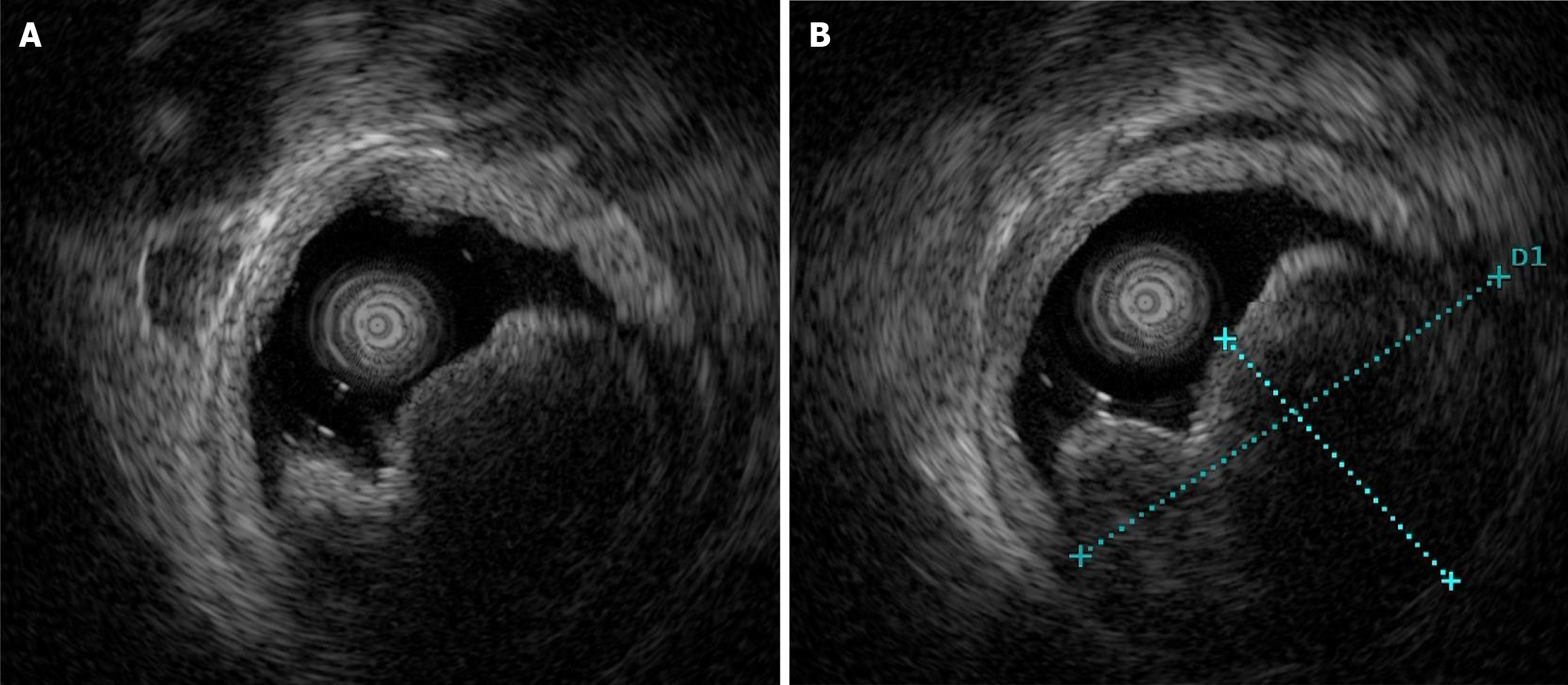
Figure 2 Ultrasonographic findings.
A: The normal hierarchy of the lesion disappeared and was replaced by a hypoechoic uniform structure involving the muscularis propria, and the serosal layer was basically complete; B: The tumor was approximately 37.1 cm × 24.5 cm in size.
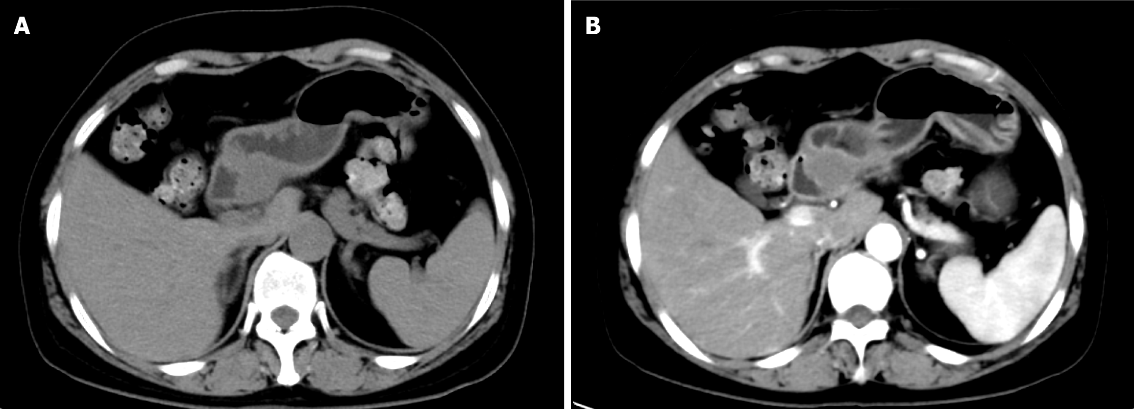
Figure 3 Computed tomography findings.
A: An oval lesion with a clear border, which had grown into the lumen and showed soft tissue density during a plain scan with a computed tomography (CT) value of 37 HU; B: The lesion showed moderate sustained enhancement during contrast enhancement with a CT value of 48 HU.
Figure 4 Magnetic resonance imaging showed an oval lesion with a clear edge in the duodenal bulb.
A: High signal on the T2-weighted magnetic resonance image; B: Equal signal on the T1-weighted magnetic resonance image; C: Enhanced scan showed that the lesion was significantly strengthened, with a small amount of low signal seen inside.
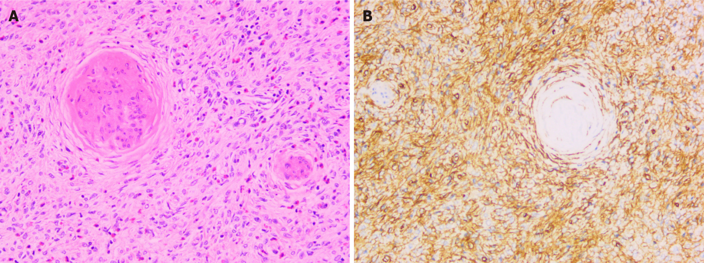
Figure 5 Pathological findings of surgical specimens.
A: Tumor cells are spindle, polygonal, arranged in bundles, mixed with eosinophils, lymphoid and plasma cells, with rare nuclear division, and involved the subserosa; B: Immunohistochemical staining revealed diffuse strongly positive CD34 with proliferating spindle cells growing "onion skin like" around small vessels.
- Citation: Zhang FM, Ning LG, Wang JJ, Zhu HT, Feng MB, Chen HT. Invasive inflammatory fibrotic polyp of the duodenum: A case report. World J Gastrointest Surg 2025; 17(8): 107558
- URL: https://www.wjgnet.com/1948-9366/full/v17/i8/107558.htm
- DOI: https://dx.doi.org/10.4240/wjgs.v17.i8.107558


