Copyright
©The Author(s) 2025.
World J Gastrointest Surg. May 27, 2025; 17(5): 105423
Published online May 27, 2025. doi: 10.4240/wjgs.v17.i5.105423
Published online May 27, 2025. doi: 10.4240/wjgs.v17.i5.105423
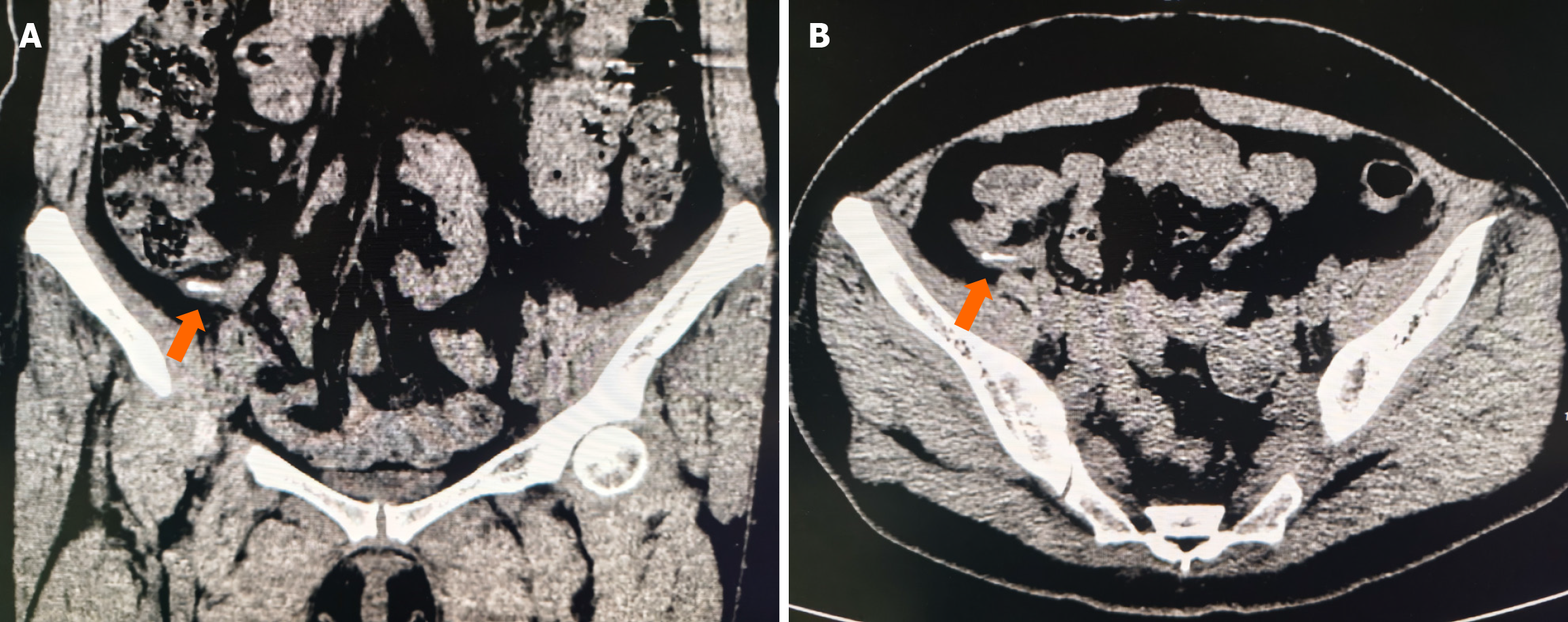
Figure 1 Abdominal computed tomography scan.
A: Coronal position; B: Cross section, both marked by orange arrows, high-density shadow in the appendiceal cavity, with slightly thickened appendiceal mesangium.
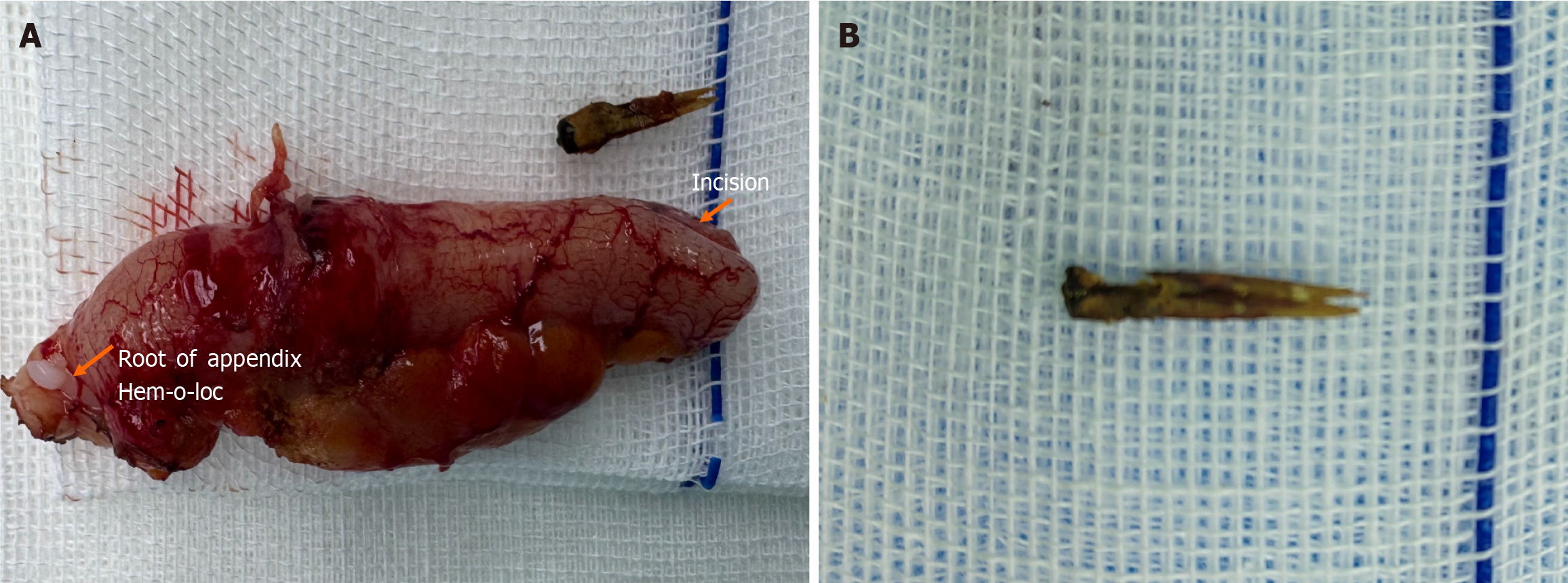
Figure 2 Surgical specimens.
A: The appendix was approximately 6 cm long, the tube diameter was normal, the serous membrane was slightly congested, the tube wall became hard, and the mesangium was slightly thickened; B: Chicken bone.
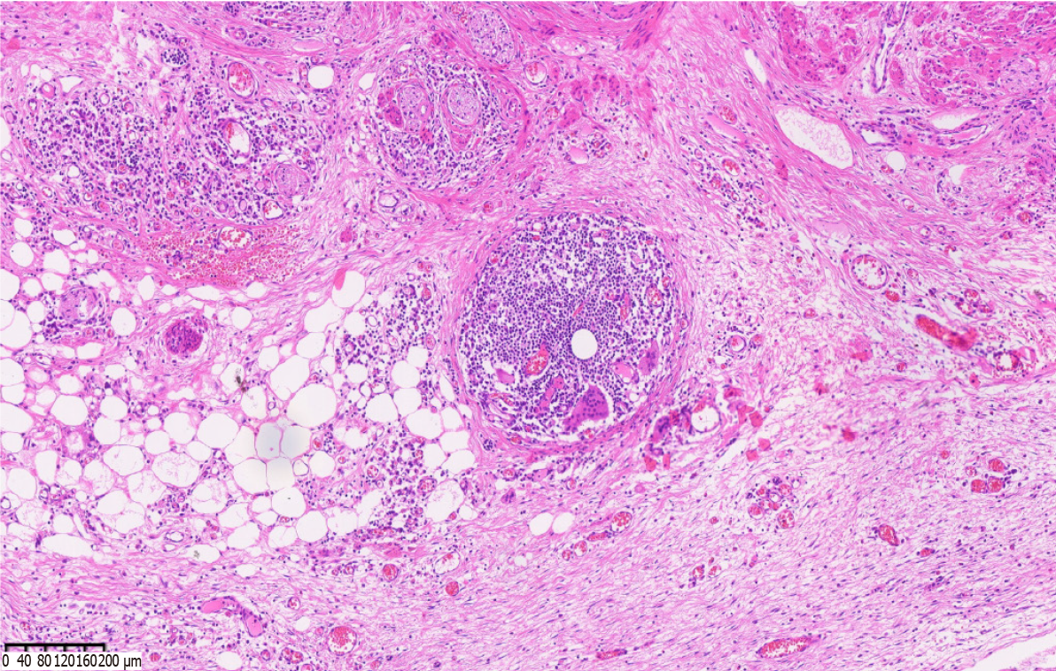
Figure 3 Postoperative histopathological examination.
Chronic appendicitis with foreign body giant cell reaction (hematoxylin and eosin, 200 ×).
- Citation: Huang T, Li SK, Wang W, Zhang R. Chronic abdominal pain caused by foreign bodies in the appendix: A case report. World J Gastrointest Surg 2025; 17(5): 105423
- URL: https://www.wjgnet.com/1948-9366/full/v17/i5/105423.htm
- DOI: https://dx.doi.org/10.4240/wjgs.v17.i5.105423


