Copyright
©The Author(s) 2025.
World J Gastrointest Surg. May 27, 2025; 17(5): 105023
Published online May 27, 2025. doi: 10.4240/wjgs.v17.i5.105023
Published online May 27, 2025. doi: 10.4240/wjgs.v17.i5.105023
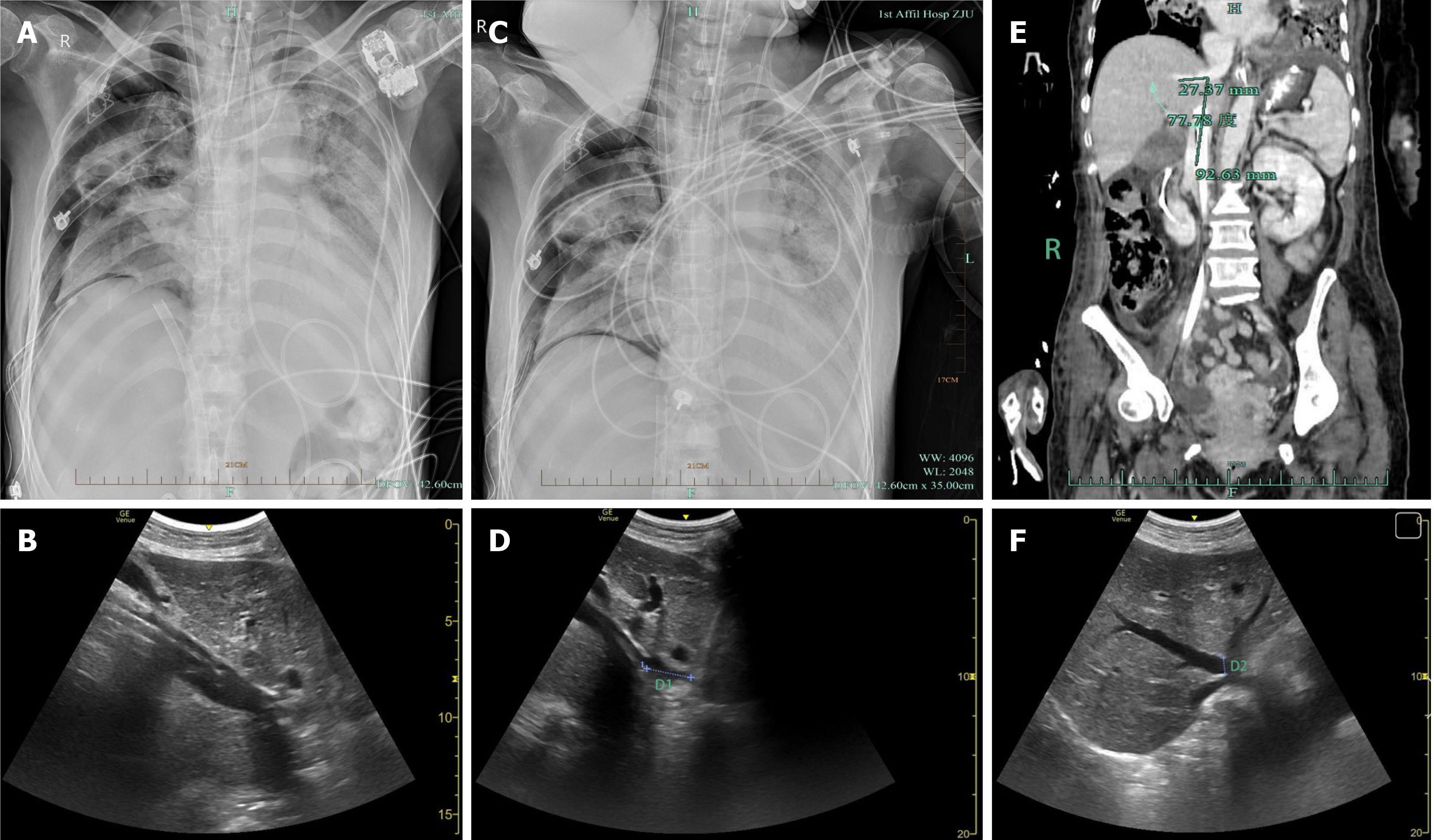
Figure 1 Computed tomography and ultrasonography radiographs during the hospital stay.
A: Chest radiography after extracorporeal membrane oxygenation (ECMO) initiation showing the ECMO venous drainage cannula tip positioned within the middle hepatic vein, along with severe bilateral pulmonary infections and a right-sided pneumothorax; B: Bedside ultrasonography confirming the ECMO venous drainage cannula tip in the middle hepatic vein following ECMO initiation; C: Chest radiography following adjustment of the ECMO venous drainage cannula, with the cannula tip now located within the inferior vena cava; D: Following adjustment under ultrasonographic guidance, the ECMO venous drainage cannula tip is positioned in the inferior vena cava at a distance of 2.71 cm from the right atrial opening (D1); E: Contrast-enhanced abdominal computed tomography angiography demonstrating an angle of 77.78° between the middle hepatic vein and the inferior vena cava; F: Ultrasonographic measurement of the middle hepatic vein opening diameter to be approximately 1.02 cm (D2).
- Citation: Li K, Pan XJ, Liu TT, Guo HY, Fang XL. Rare complication of extracorporeal membrane oxygenation cannula misplacement into the hepatic vein: A case report. World J Gastrointest Surg 2025; 17(5): 105023
- URL: https://www.wjgnet.com/1948-9366/full/v17/i5/105023.htm
- DOI: https://dx.doi.org/10.4240/wjgs.v17.i5.105023


