Copyright
©The Author(s) 2025.
World J Gastrointest Surg. May 27, 2025; 17(5): 103078
Published online May 27, 2025. doi: 10.4240/wjgs.v17.i5.103078
Published online May 27, 2025. doi: 10.4240/wjgs.v17.i5.103078
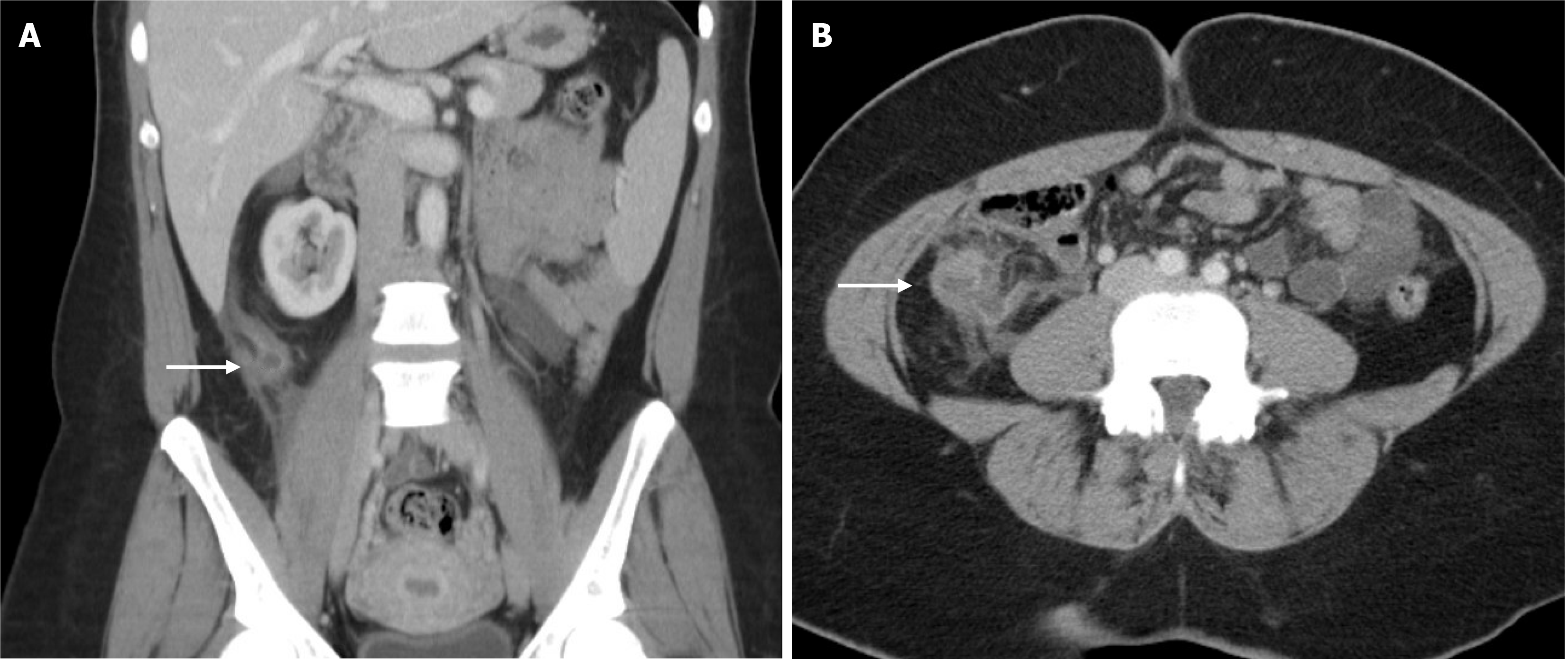
Figure 1 Computed tomography images of the patient at the initial presentation.
A: Coronal computed tomography (CT) image with an arrow indicating a locule of fluid; B: Axial CT image with an arrow indicating a locule of fluid and surrounding inflammation.
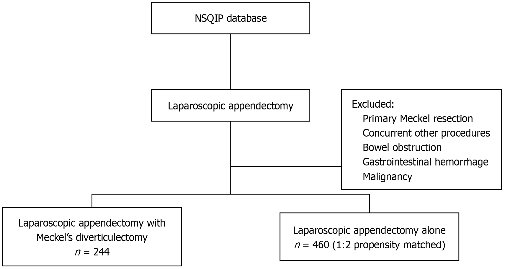
Figure 2
Inclusion and exclusion criteria.
- Citation: Nguyen SHT, Wheelwright M, Vakayil V, Meshram P, O’Donnell R, Harmon JV. Concomitant resection of Meckel diverticulum during laparoscopic appendectomy: Retrospective propensity-matched ACS-NSQIP study and a case report. World J Gastrointest Surg 2025; 17(5): 103078
- URL: https://www.wjgnet.com/1948-9366/full/v17/i5/103078.htm
- DOI: https://dx.doi.org/10.4240/wjgs.v17.i5.103078


