Copyright
©The Author(s) 2025.
World J Gastrointest Surg. Apr 27, 2025; 17(4): 104726
Published online Apr 27, 2025. doi: 10.4240/wjgs.v17.i4.104726
Published online Apr 27, 2025. doi: 10.4240/wjgs.v17.i4.104726
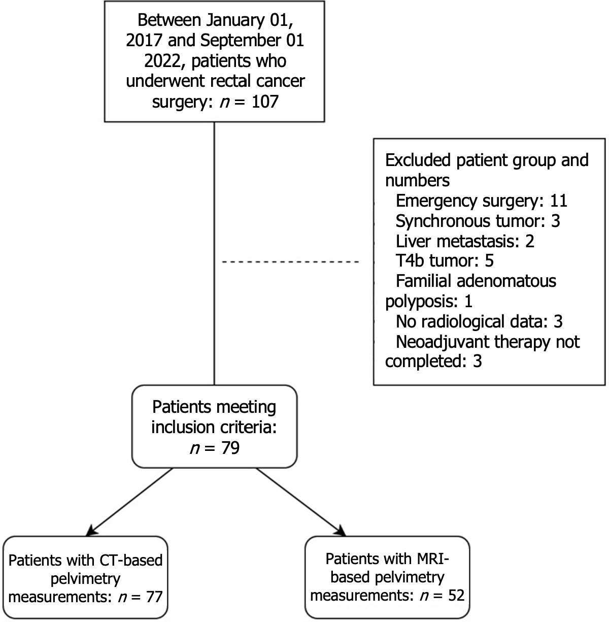
Figure 1 Flowchart of research methodology.
CT: Computed tomography.
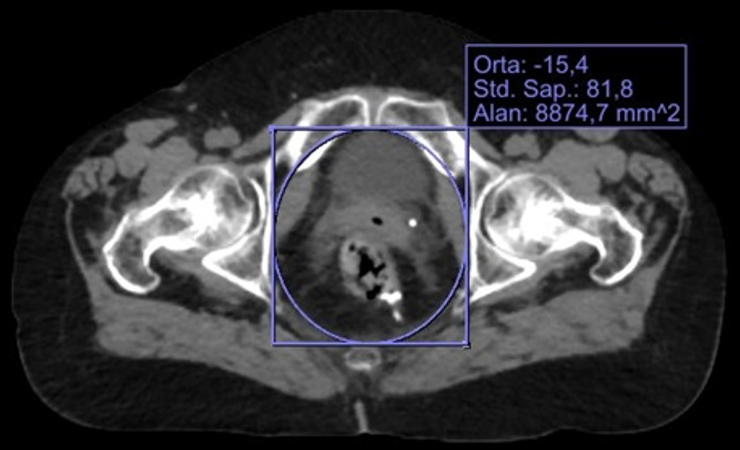
Figure 2 Pelvic inlet area is calculated using the Picture Archiving and Communication System.
Figure 3 Magnetic resonance imaging-based calculation of the pelvic cavity index.
A: Measurement of pelvic inlet diameter and pelvic depth in sagittal section using magnetic resonance imaging; B: Measurement of interspinous distance using magnetic resonance imaging [pelvic cavity index: (a.c/b)].
- Citation: Ay OF, Firat D, Özçetin B, Ocakoglu G, Ozcan SGG, Bakır Ş, Ocak B, Taşkin AK. Role of pelvimetry in predicting surgical outcomes and morbidity in rectal cancer surgery: A retrospective analysis. World J Gastrointest Surg 2025; 17(4): 104726
- URL: https://www.wjgnet.com/1948-9366/full/v17/i4/104726.htm
- DOI: https://dx.doi.org/10.4240/wjgs.v17.i4.104726


