Copyright
©The Author(s) 2025.
World J Gastrointest Surg. Feb 27, 2025; 17(2): 99423
Published online Feb 27, 2025. doi: 10.4240/wjgs.v17.i2.99423
Published online Feb 27, 2025. doi: 10.4240/wjgs.v17.i2.99423
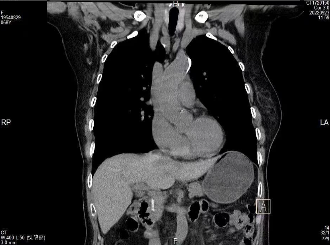
Figure 1
Computed tomography coronal view shows a strip of high-density shadow in the common bile duct.
Figure 2 Magnetic resonance cholangiopancreatography showing a low-signal foreign body in the common bile duct (the external shape is similar to a Hem-o-lok clip).
A-C: High-density shadow in the common bile duct.
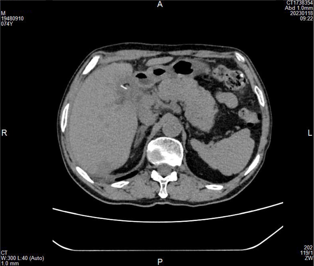
Figure 3
A high-density strip shadow can be seen near the T-tube sinus.
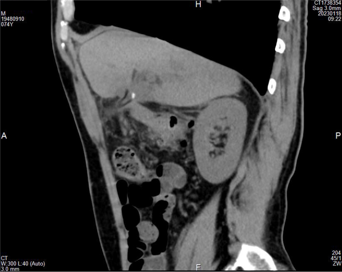
Figure 4
High-density shadows can be seen.
Figure 5
Endoscopic retrograde cholangiopancreatography shows a stone in the common bile duct centered on the Hem-o-lok clip.
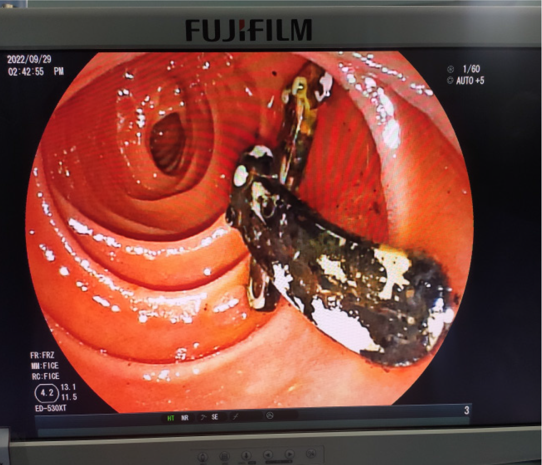
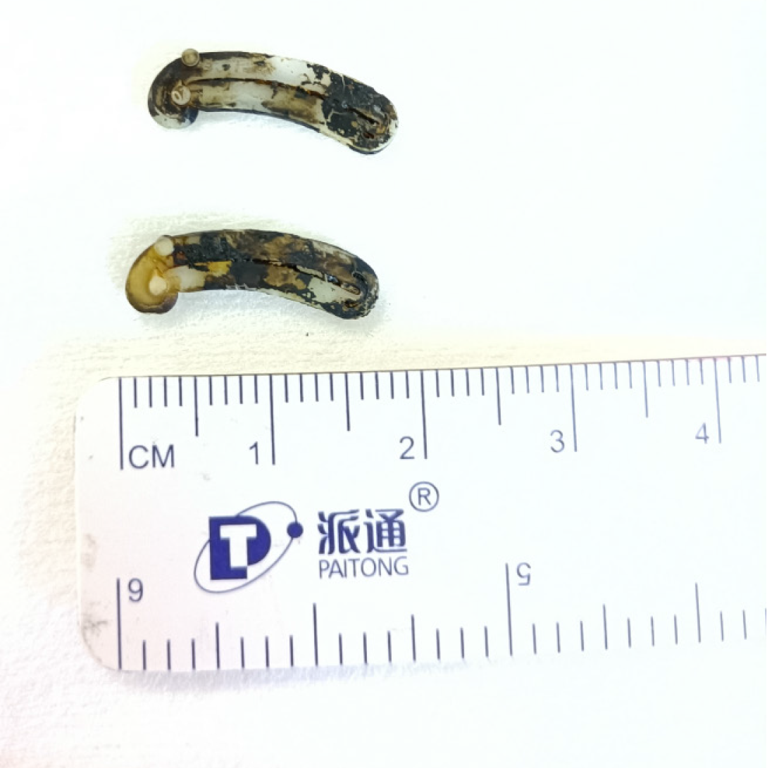
Figure 6
Stone fragments and two large Hem-o-lok clips were removed using a stone balloon.
- Citation: Huang YZ, Lin YY, Xie JP, Deng G, Tang D. Clip-stone and T clip-sinus post laparoscopic biliary surgery: Two case reports and review of the literature. World J Gastrointest Surg 2025; 17(2): 99423
- URL: https://www.wjgnet.com/1948-9366/full/v17/i2/99423.htm
- DOI: https://dx.doi.org/10.4240/wjgs.v17.i2.99423


