Copyright
©The Author(s) 2025.
World J Gastrointest Surg. Feb 27, 2025; 17(2): 101239
Published online Feb 27, 2025. doi: 10.4240/wjgs.v17.i2.101239
Published online Feb 27, 2025. doi: 10.4240/wjgs.v17.i2.101239
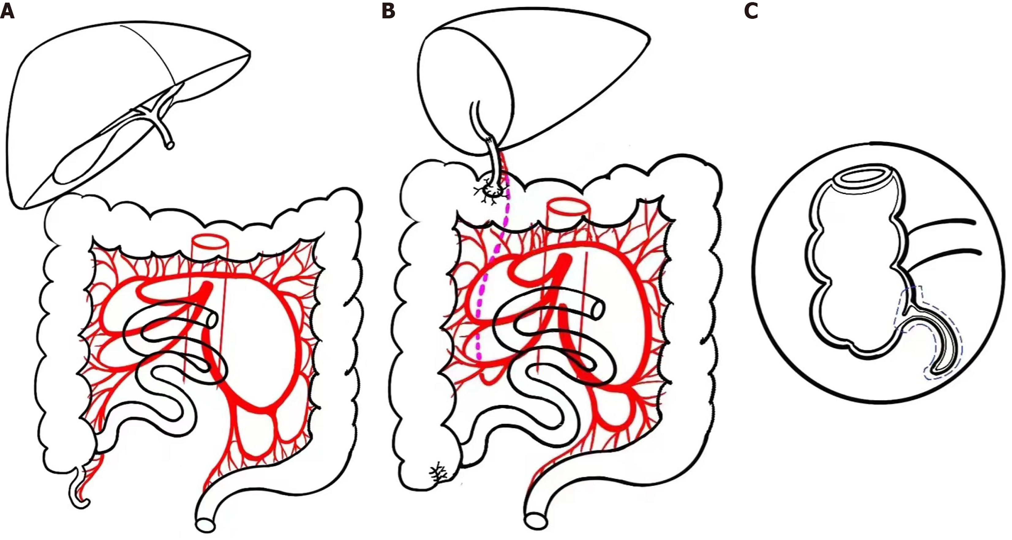
Figure 1 Surgical technique schematic diagram.
A: Excision of the diseased liver; B: Biliary reconstruction; C: Excision range of appendix.
Figure 2 Imaging examination.
A: Upper abdomen computerized tomography performed 4 months after liver transplantation (LT) operation; B: Upper abdomen computerized tomography performed 10 months after LT operation; C: Radionuclide cholangiography performed 10 months after LT operation (upper left corner: Cholangiography 45 minutes after the injection of radionuclide, nuclide concentrated in intrahepatic bile duct; upper right corner: Cholangiography 1.5 hours after the injection of radionuclide, nuclide concentrated in anastomosed colon; lower left corner: Cholangiography 4 hours after the injection of radionuclide, nuclide concentrated in transverse colon; lower right corner: Cholangiography 5 hours after the injection of radionuclide, nuclide concentrated in the whole colon).
- Citation: Song JQ, Zhou T, Luo Y, Liu Y. Internal biliary diversion using appendix during liver transplantation for progressive familial intrahepatic cholestasis type 1: A case report. World J Gastrointest Surg 2025; 17(2): 101239
- URL: https://www.wjgnet.com/1948-9366/full/v17/i2/101239.htm
- DOI: https://dx.doi.org/10.4240/wjgs.v17.i2.101239


