Copyright
©The Author(s) 2025.
World J Gastrointest Surg. Jan 27, 2025; 17(1): 99758
Published online Jan 27, 2025. doi: 10.4240/wjgs.v17.i1.99758
Published online Jan 27, 2025. doi: 10.4240/wjgs.v17.i1.99758
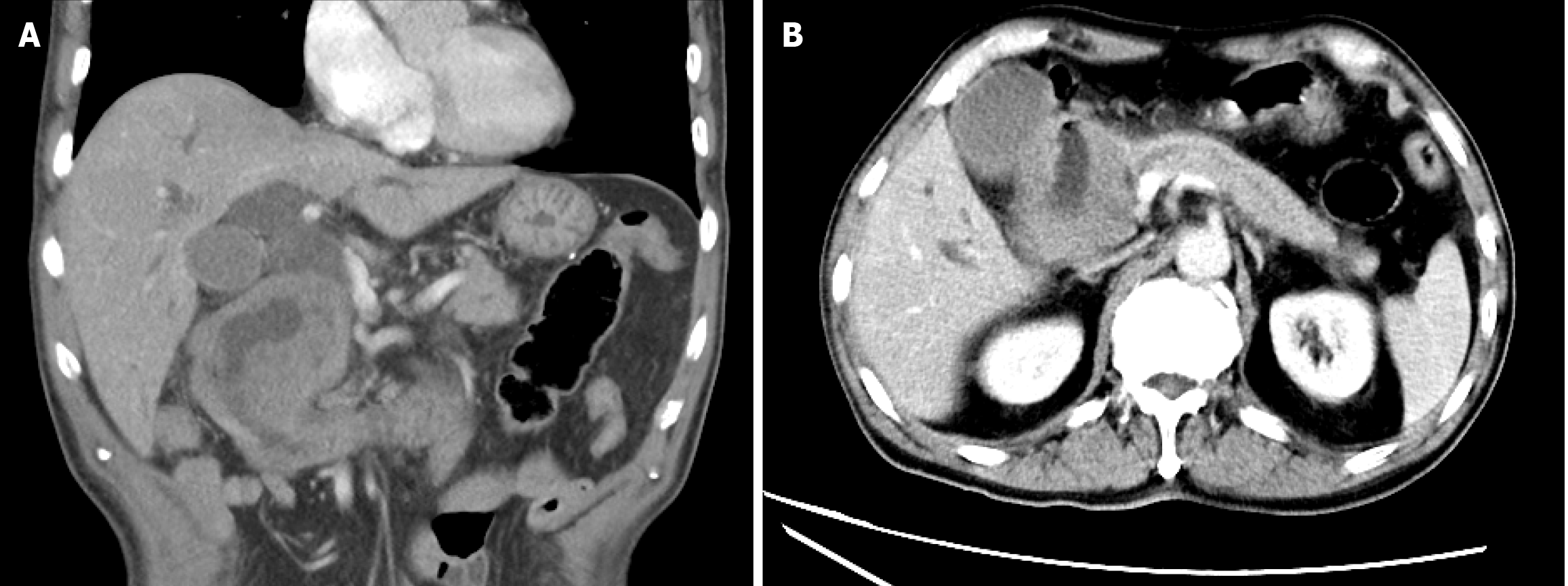
Figure 1 Abdominal computed tomography scan.
A: Heterogeneous thickening of the descending duodenum with a 7.6 cm × 7.4 cm × 7.4 cm low-density mass, luminal narrowing, and dilation of the intrahepatic and extrahepatic bile ducts; B: Gallbladder enlargement and main pancreatic duct dilatation.
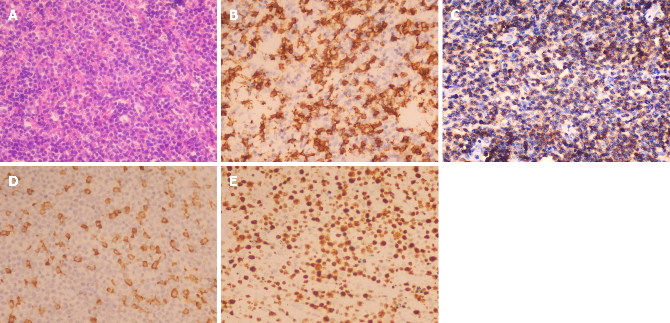
Figure 2 Histopathological and immunohistochemistry examination.
A: Large atypical lymphoid cells were scattered among numerous small reactive lymphocytes and, less often, histiocytes (hematoxylin-eosin, × 40); B: CD20-positive large, atypical B cells (× 40); C: Numerous CD3-positive small T cells in the background (× 40); D: CD163-positive histiocytes in the background (× 40); E: Ki-67 expression in neoplastic cells (× 40).
- Citation: Chen XY, Yang JY, Chen YH, Liu AN, Wu SS, Ji Zhi SN, Zheng SM. Primary duodenal T/histiocyte-rich large B-cell lymphoma complicated with obstructive jaundice: A case report and review of literature. World J Gastrointest Surg 2025; 17(1): 99758
- URL: https://www.wjgnet.com/1948-9366/full/v17/i1/99758.htm
- DOI: https://dx.doi.org/10.4240/wjgs.v17.i1.99758


