Copyright
©The Author(s) 2025.
World J Gastrointest Surg. Jan 27, 2025; 17(1): 99752
Published online Jan 27, 2025. doi: 10.4240/wjgs.v17.i1.99752
Published online Jan 27, 2025. doi: 10.4240/wjgs.v17.i1.99752
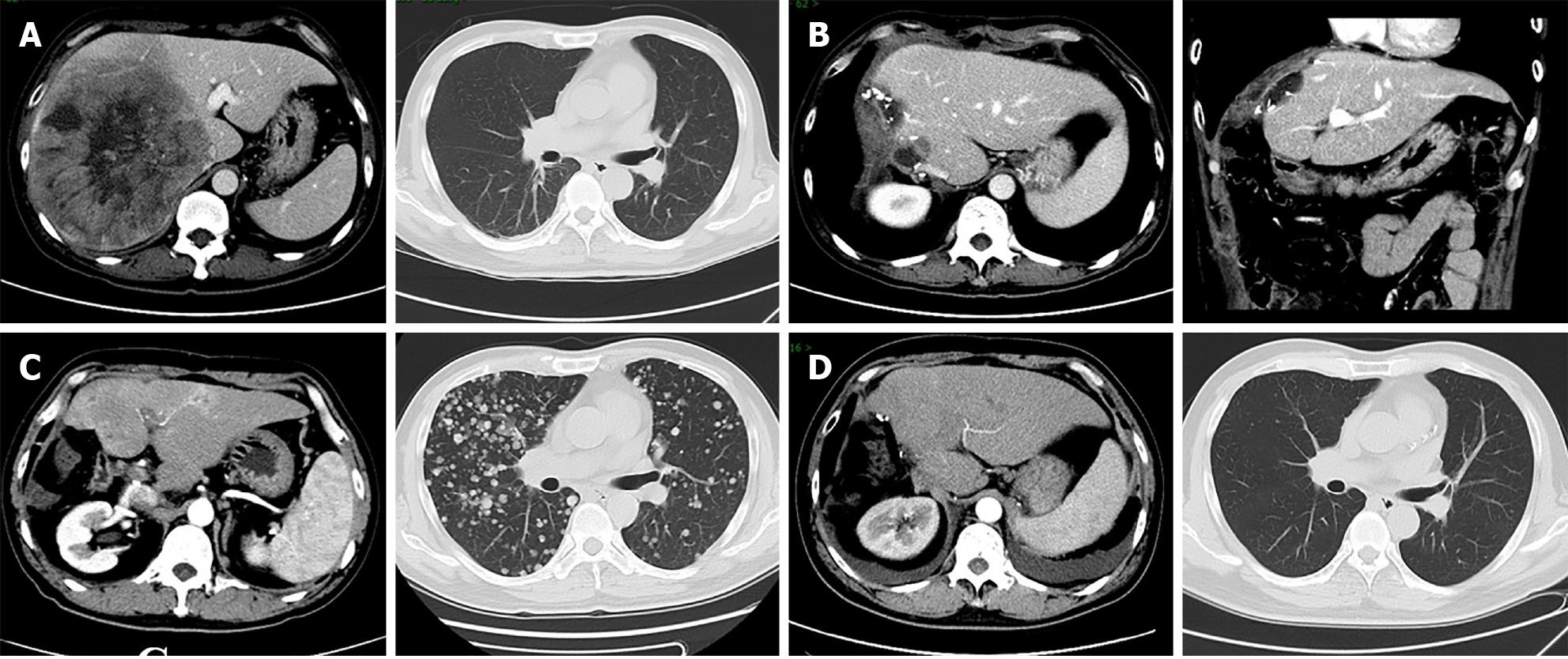
Figure 1 Contrast-enhanced abdominal computed tomography and contrast-free thoracic computed tomography.
A: Computed tomography (CT) scans showed a super-giant tumor 152 mm × 171 mm in size in the right liver lobe, without lung metastasis; B: The postoperative CT scans showed that hepatocellular carcinoma was removed; C: CT scans showed multiple liver recurrences and lung metastasis in December 2019; D: CT scans showed complete remission of liver recurrence and lung metastasis on May 12, 2020.
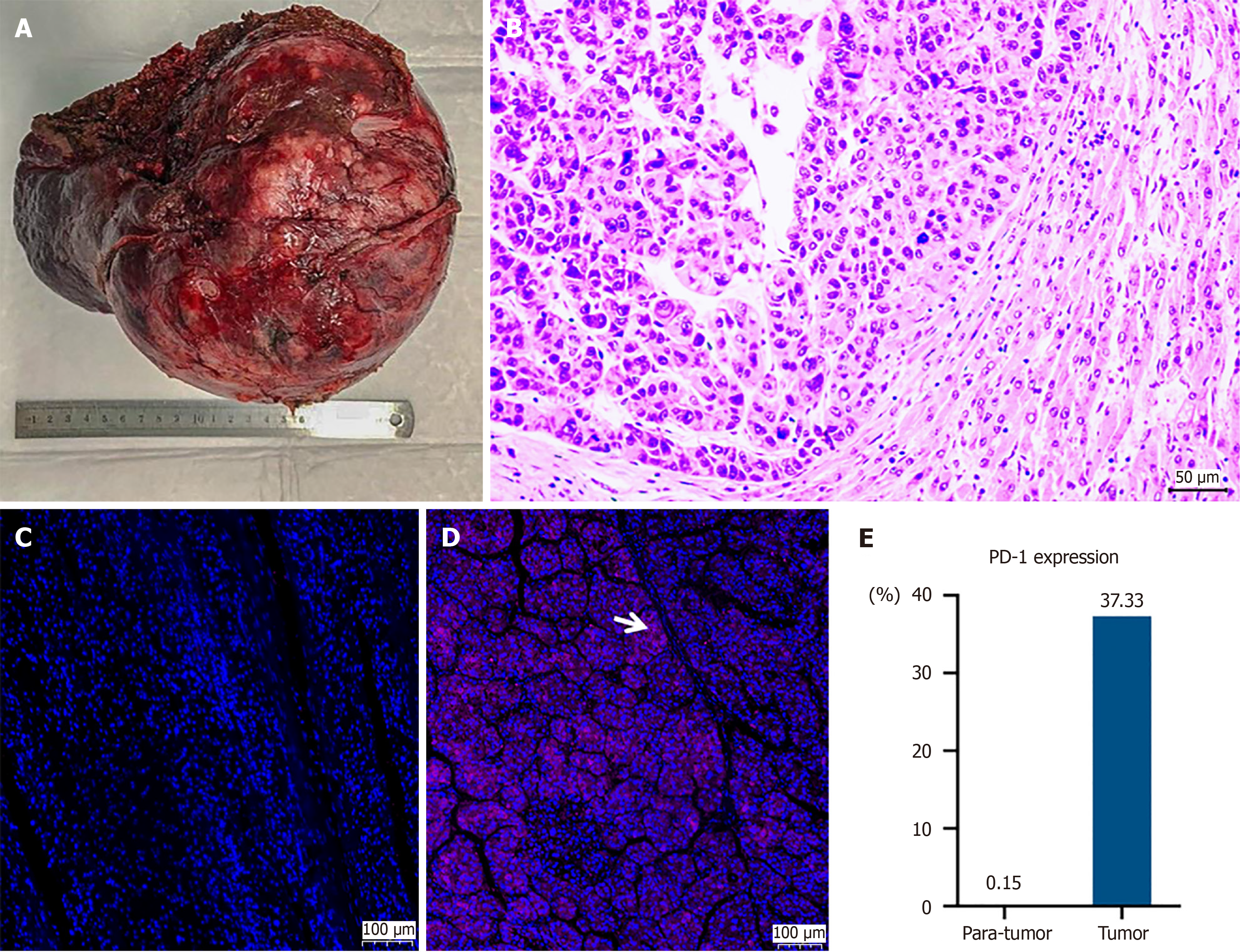
Figure 2 Histopathology of the resected liver tumor.
A: The specimen showed a giant hepatic tumor over 170 mm in size; B: Hematoxylin and eosin staining of hepatocellular carcinoma revealed that the tumor cells were arranged in a nest-like cluster at the junction with normal liver tissue, and the nuclear was large and deeply stained with atypical morphology (200 × magnification); C-E: Immunofluorescence staining of programmed cell death receptor 1 in tumor tissue showed more positive findings than in para-carcinoma tissues. The arrow indicates positive. PD-1: Programmed cell death receptor 1.
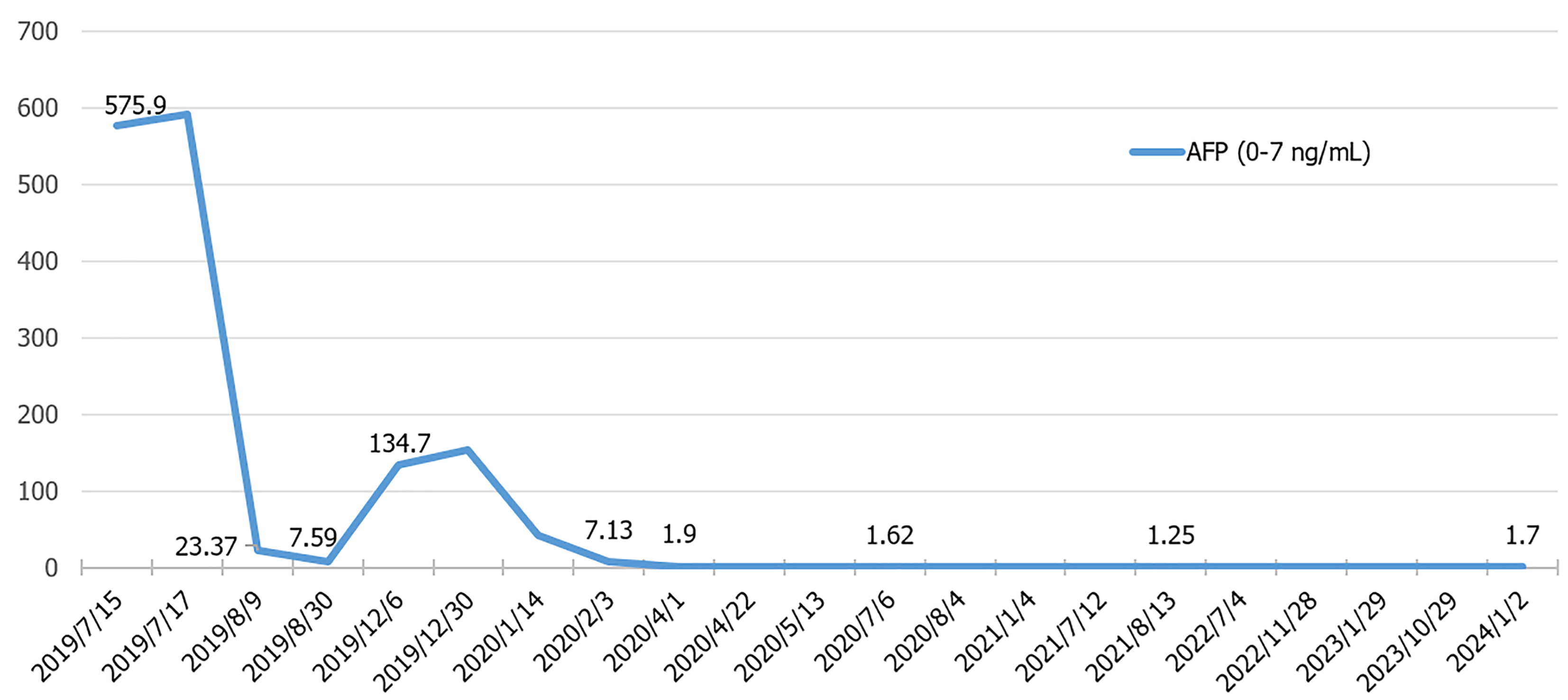
Figure 3 Dynamic change in α-fetoprotein during the entire treatment period.
AFP: α-fetoprotein.
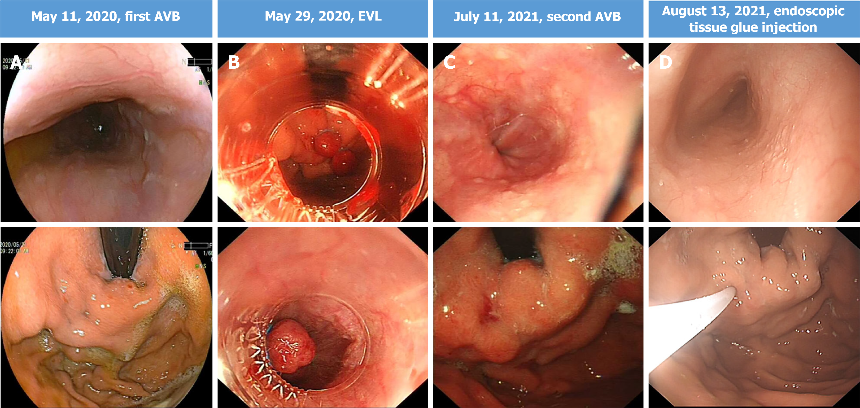
Figure 4 Endoscopy examinations.
After two episodes of gastrointestinal bleeding, upper gastrointestinal endoscopy revealed mild esophagogastric varices without red color signs. The patient was subsequently treated with esophageal variceal ligation and endoscopic tissue glue injection. A: The endoscopy finds the first acute esophageal variceal bleeding on May 11, 2020; B: The patient accepted endoscopic variceal ligation treatment on May 29, 2020; C: The endoscopy found the gastric variceal bleeding without esophageal varices on July 11, 2021; D: The patient accepted endoscopic tissue glue injection treatment on August 13, 2021. AVB: Acute variceal bleeding; EVL: Endoscopic variceal ligation.
Figure 5 The entire treatment timeline.
TACE: Transcatheter arterial chemoembolization; HCC: Hepatocellular carcinoma; AVB: Acute variceal bleeding; EVL: Endoscopic variceal ligation.
- Citation: Zheng XQ, Sun LB, Jin WJ, Liu H, Song WY, Xu H, Wu JS, Wang XJ, Gou CY, Ding HG. Five-year complete remission of super-giant hepatocellular carcinoma with hepatectomy followed by sorafenib plus camrelizumab: A case report. World J Gastrointest Surg 2025; 17(1): 99752
- URL: https://www.wjgnet.com/1948-9366/full/v17/i1/99752.htm
- DOI: https://dx.doi.org/10.4240/wjgs.v17.i1.99752


