Copyright
©The Author(s) 2025.
World J Gastrointest Surg. Jan 27, 2025; 17(1): 98283
Published online Jan 27, 2025. doi: 10.4240/wjgs.v17.i1.98283
Published online Jan 27, 2025. doi: 10.4240/wjgs.v17.i1.98283
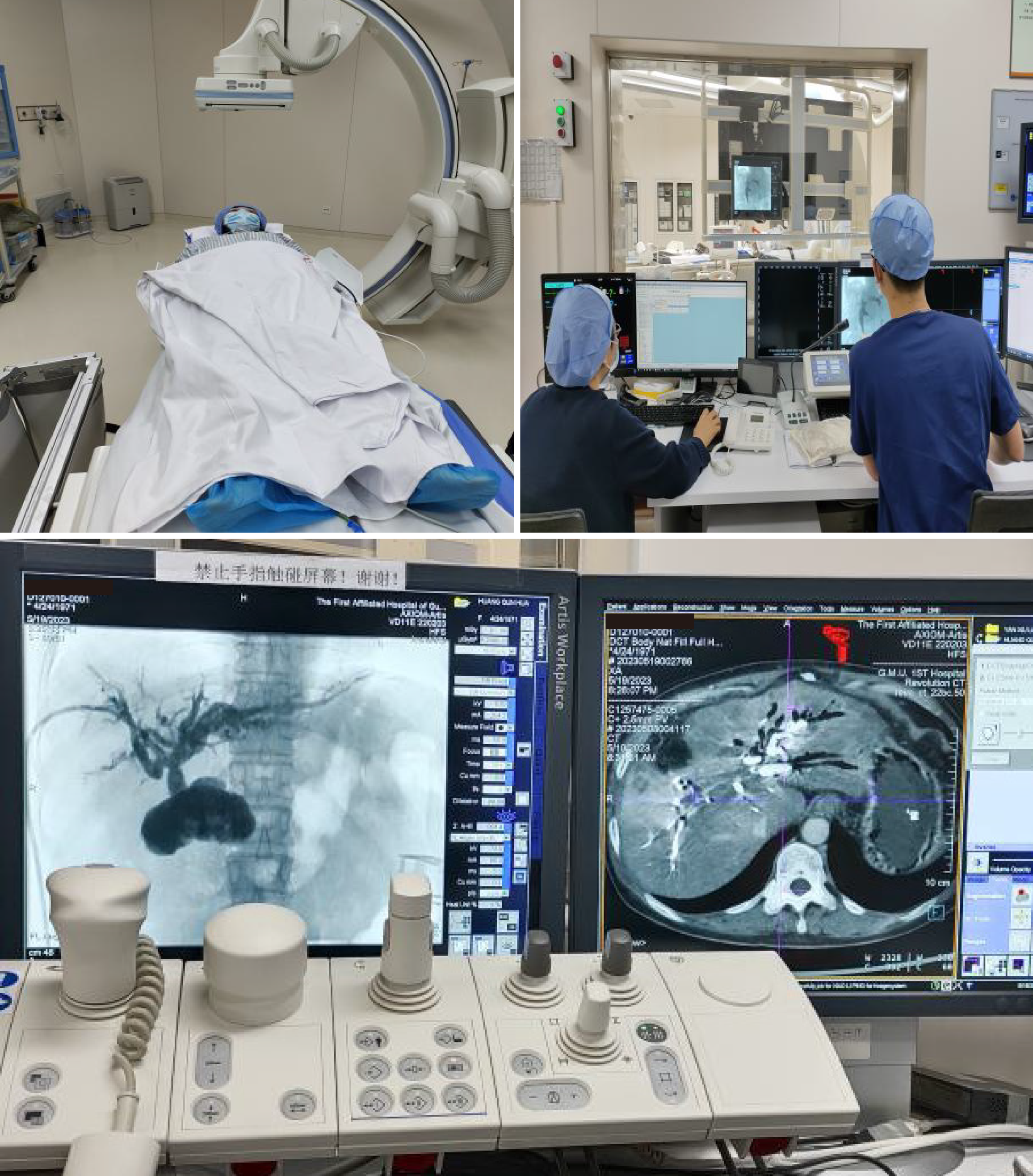
Figure 1
Process of DynaCT biliary soft tissue reconstruction technology.
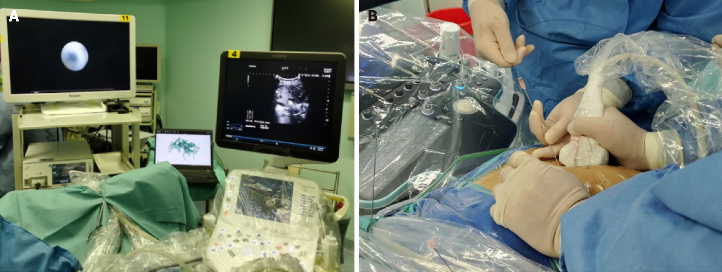
Figure 2 Three-dimensional model combined with real-time B-ultrasonic navigation.
A: Combination of three-dimensional reconstruction and B-ultrasound for guiding one-step percutaneous transhepatic cholangioscopic lithotripsy (PTCSL); B: Using the percutaneous transhepatic one-step biliary fistulation technique for one-step PTCSL.
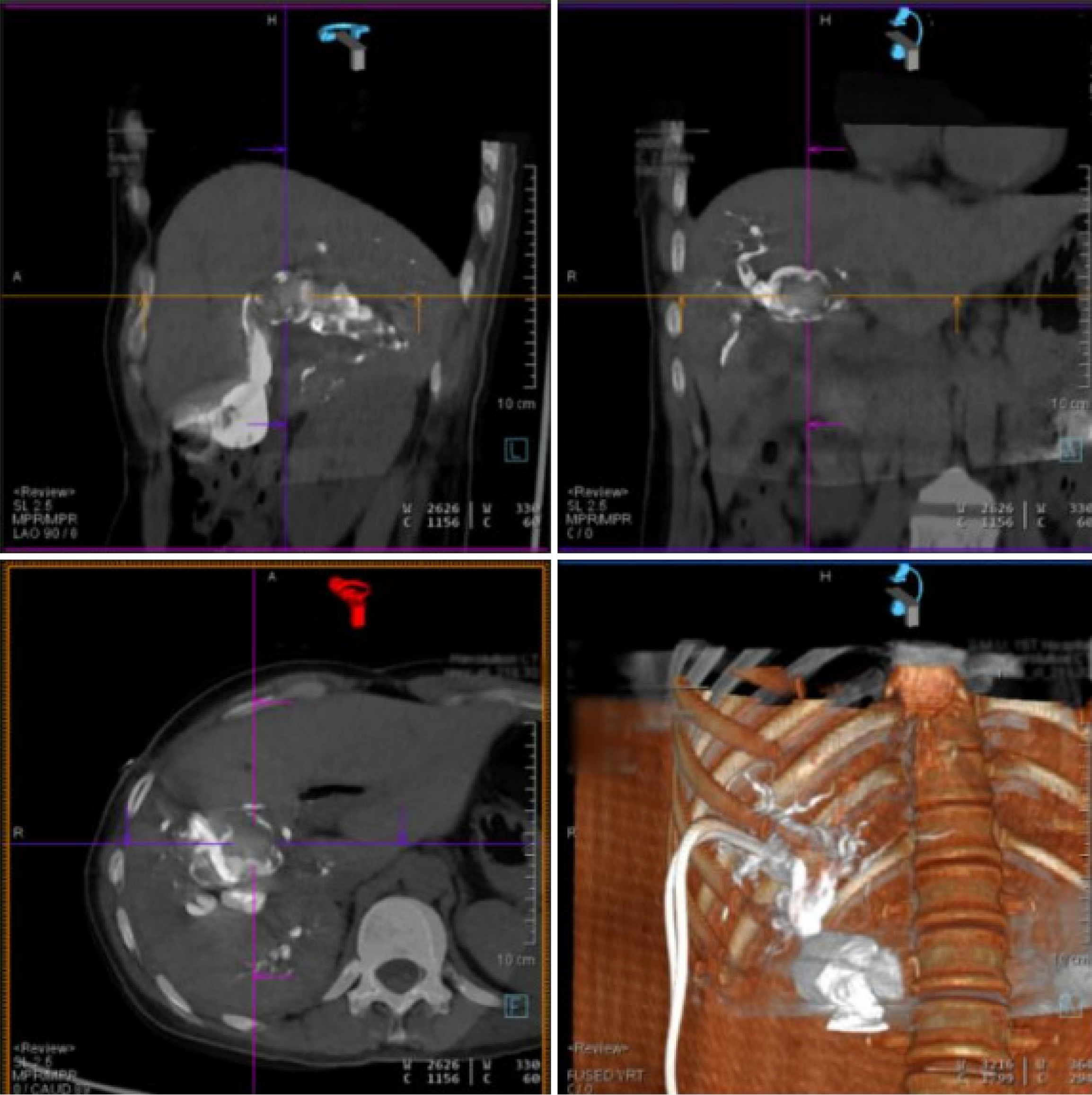
Figure 3
DynaCT soft tissue reconstruction of the biliary tract.
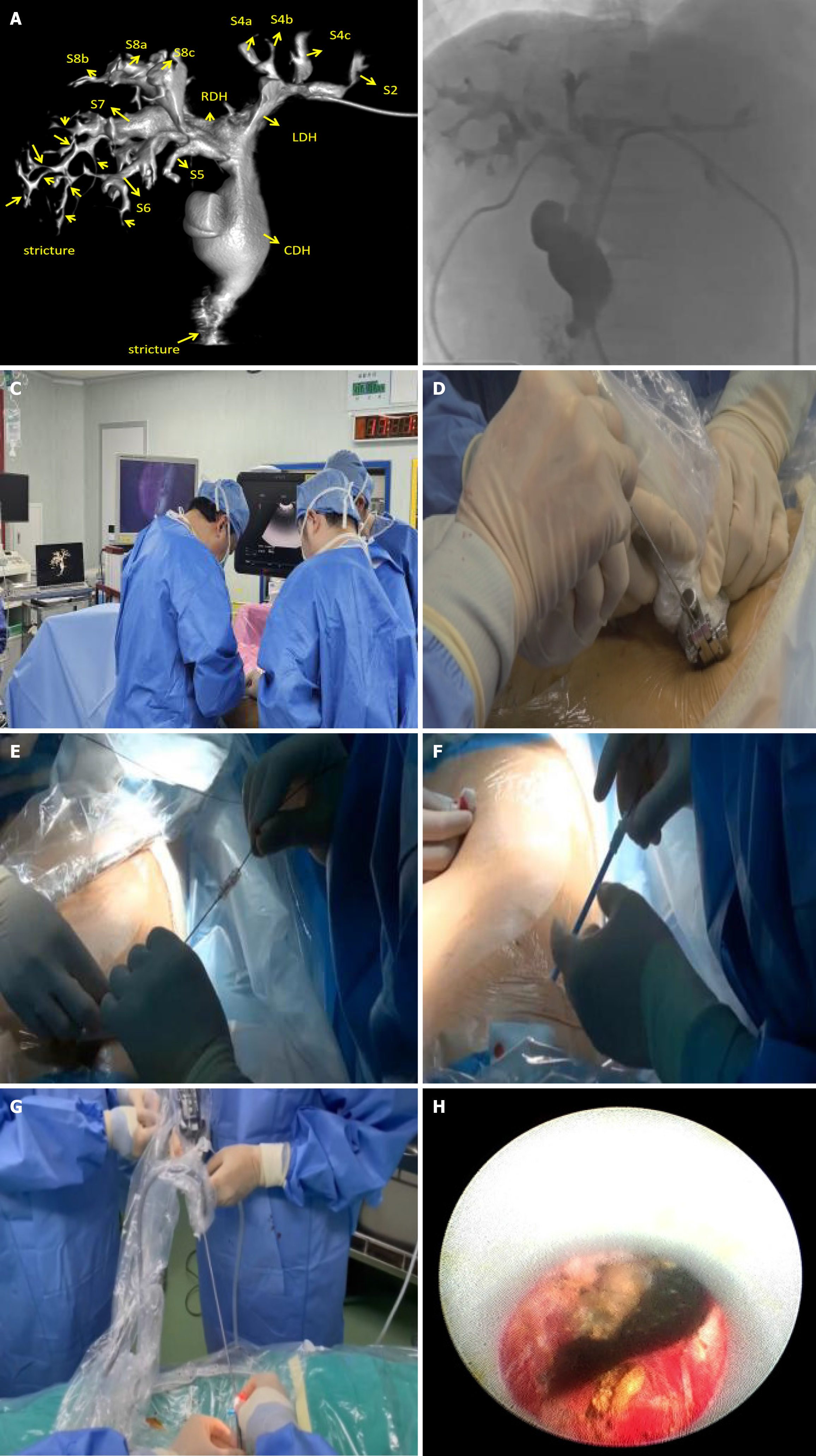
Figure 4 DynaCT-guided surgical procedure.
A: The digital reconstruction model of DynaCT three-dimensional (3D) reconstruction can realistically reproduce the sub-segments of the bile duct by measurements and 3D visualisation of the rotation, depicting the location and extent of the stricture; B: T-tube angiography image of the same patient with visible contrast agent; C: DynaCT soft tissue reconstruction image of the biliary tract and intraoperative ultrasound-guided biliary puncture; D: Biliary puncture; E: Biliary puncture with 18G needle and 0.035 inch hydrophilic guidewire; F: Fistula dilation with fascial dilators from 8 Fr to 14 Fr sequentially; G: Stone fragmentation and clearance with A 12 Fr rigid nephroscope; H: Choledochoscopic removal of stones.
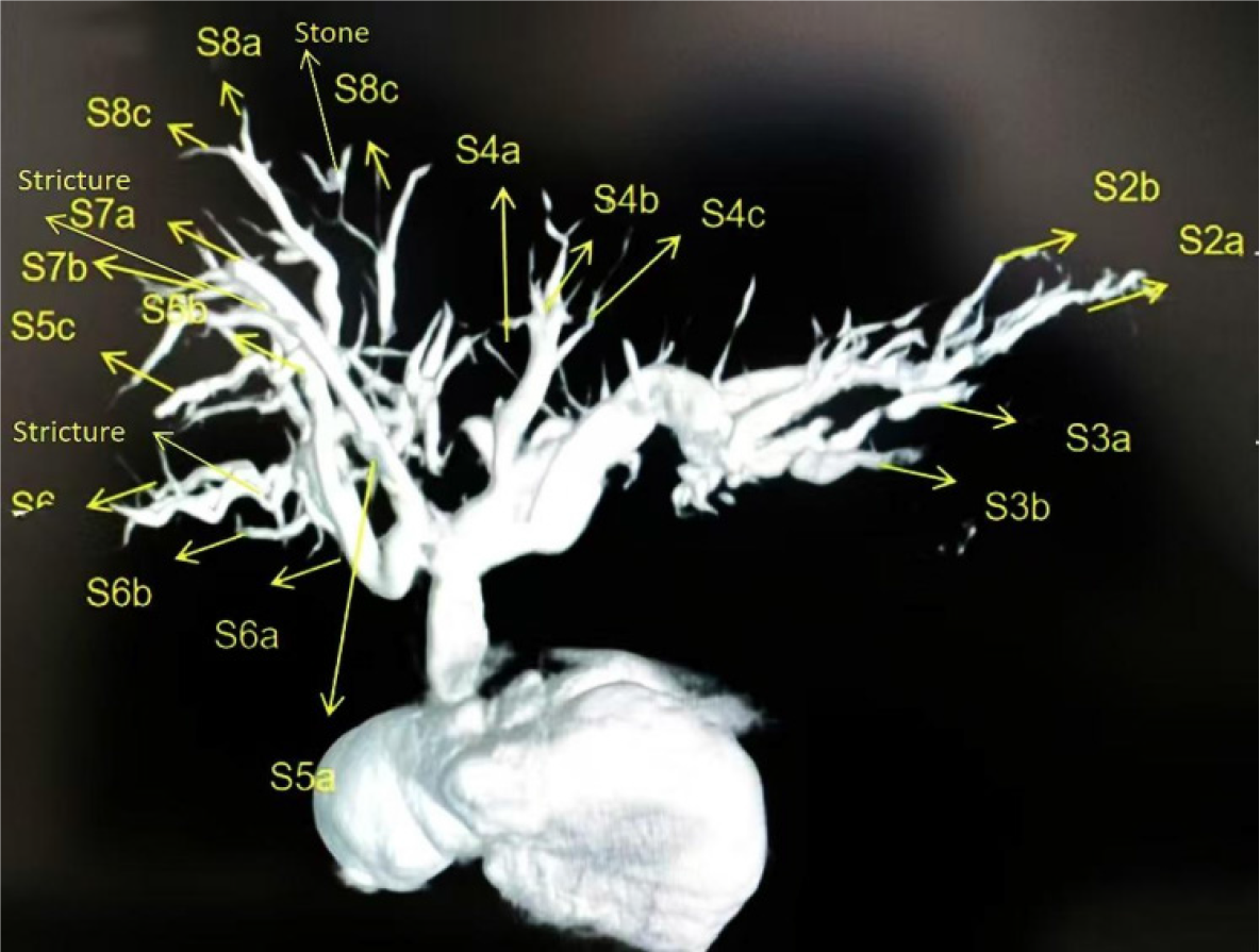
Figure 5
Digital DynaCT biliary soft tissue reconstruction of biliary branches, biliary strictures, and stones.
- Citation: Ye YQ, Li PH, Ding ZW, Zhang SF, Li RQ, Cao YW. Application of DynaCT biliary soft tissue reconstruction technology in diagnosis and treatment of hepatolithiasis. World J Gastrointest Surg 2025; 17(1): 98283
- URL: https://www.wjgnet.com/1948-9366/full/v17/i1/98283.htm
- DOI: https://dx.doi.org/10.4240/wjgs.v17.i1.98283


