Copyright
©The Author(s) 2025.
World J Gastrointest Surg. Jan 27, 2025; 17(1): 100119
Published online Jan 27, 2025. doi: 10.4240/wjgs.v17.i1.100119
Published online Jan 27, 2025. doi: 10.4240/wjgs.v17.i1.100119
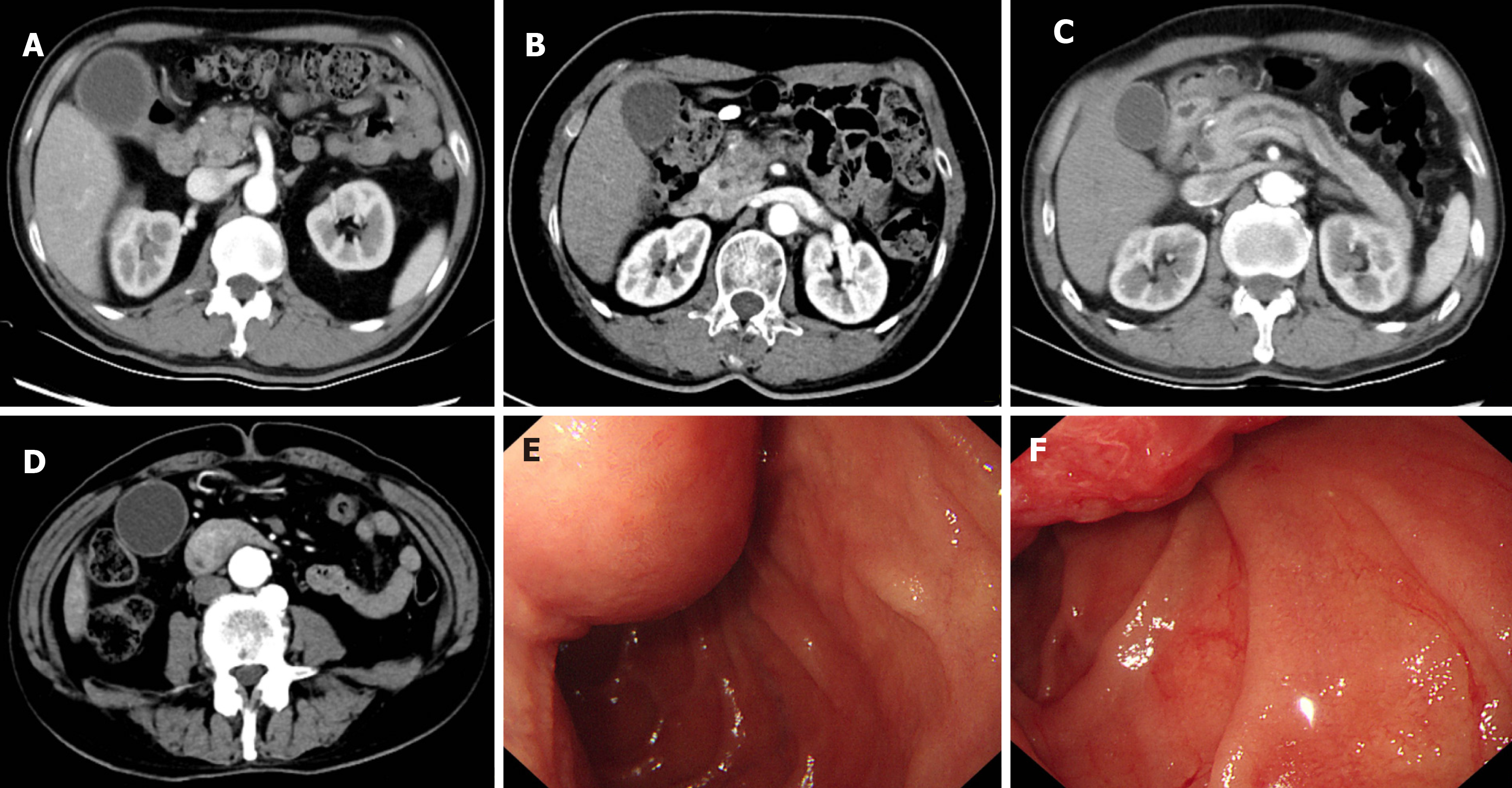
Figure 1 Computed tomography images of Cases 1-4 and endoscopy image of Case 4.
A: Computed tomography (CT) image of a 70-year-old male patient who was diagnosed with pancreatic ductal adenocarcinoma (PDAC) (Case 1); B: CT image of a 65-year-old female patient who had PDAC (Case 2); C: CT image of a 69-year-old male patient who was diagnosed with chronic pancreatitis (Case 3); D: CT image of a 71-year-old man who had periampullary adenocarcinoma (Case 4); E and F: Endoscopy images of the ampullary adenocarcinoma in Case 4.
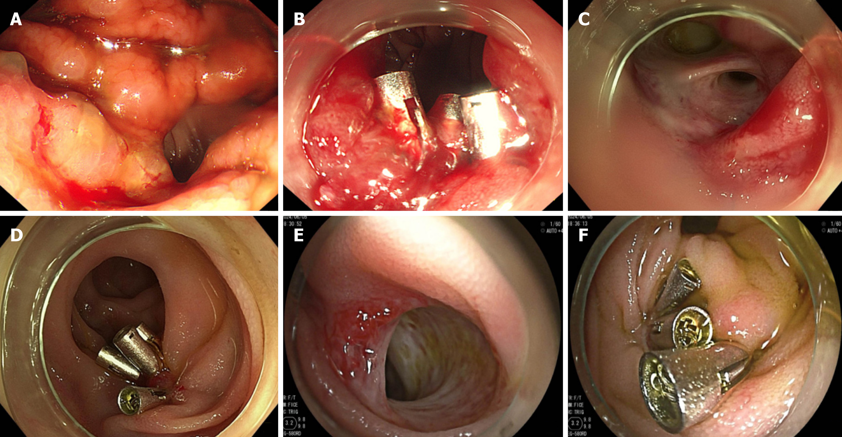
Figure 2 Endoscopy images of Cases 1 and 2.
A: Esophagogastroduodenoscopy (EGD) showed gastrointestinal anastomosis bleeding (Forrester Ib) in Case 1; B: He received three endoscopic hemostatic clips and the bleeding was stopped; C: Second EGD showed an oozing bleeding spot at his bile duct-jejunum anastomosis (Forrester Ib); D: The patient received five endoscopic hemostatic clips and hemostasis was successful; E: EGD showed vascular malformation exposed at the bile duct-jejunum anastomosis in Case 2; F: Five endoscopic hemostatic clips were applied to prevent future bleeding.
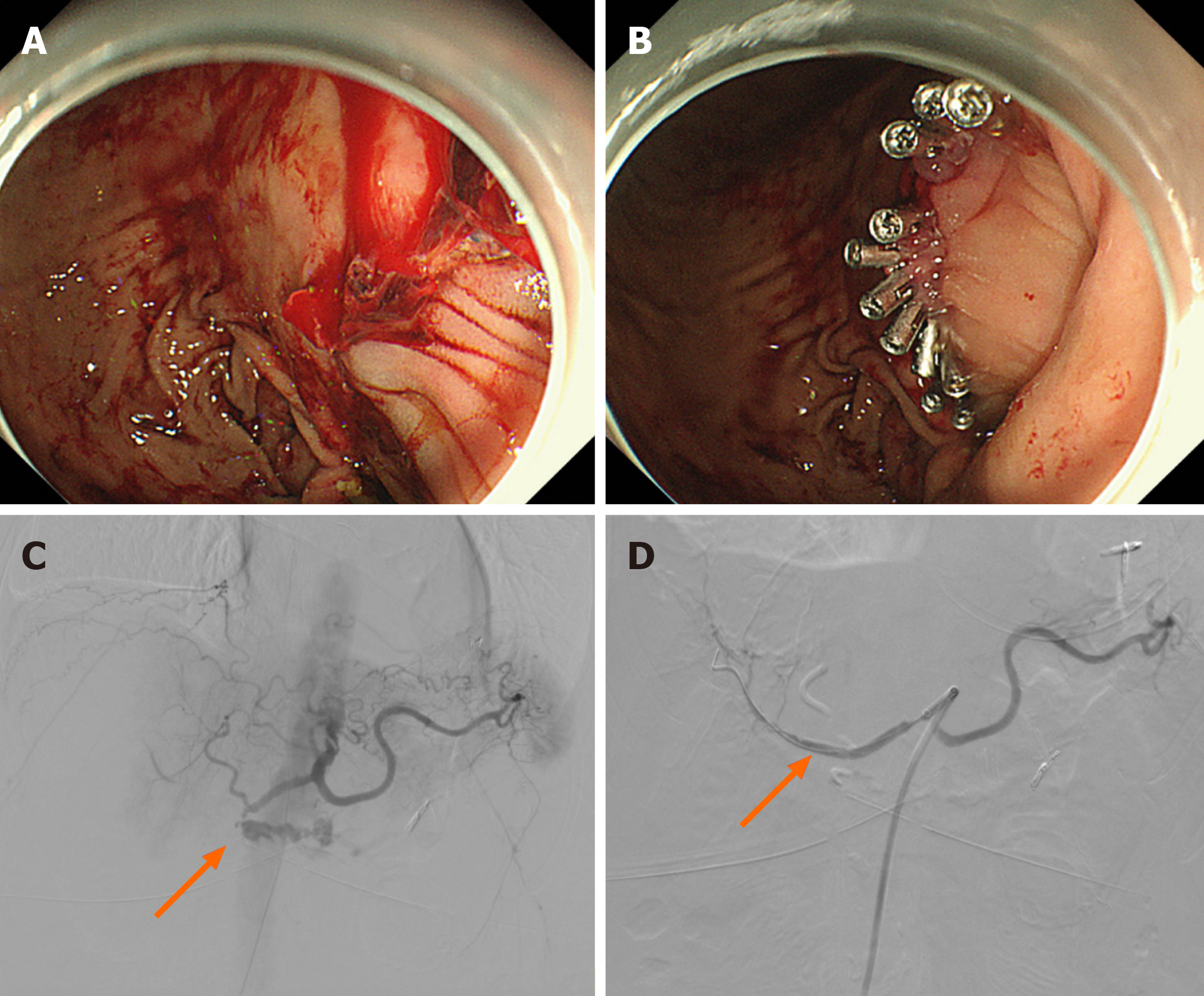
Figure 3 Endoscopy and angiography images of Cases 3 and 4.
A: Esophagogastroduodenoscopy (EGD) showed gastrointestinal anastomosis bleeding (Forrester Ib) in Case 3; B: He received 11 endoscopic hemostatic clips and the bleeding was stopped; C: Angiography showed a bleeding spot of gastroduodenal artery (orange arrow) in Case 4; D: A covered stent was placed and the bleeding was stopped temporarily (orange arrow).
- Citation: Liu ZJ, Hong JY, Zhang C, She J, Zhai HH. Gastrointestinal bleeding after pancreatoduodenectomy: Report of four cases. World J Gastrointest Surg 2025; 17(1): 100119
- URL: https://www.wjgnet.com/1948-9366/full/v17/i1/100119.htm
- DOI: https://dx.doi.org/10.4240/wjgs.v17.i1.100119


