Copyright
©The Author(s) 2024.
World J Gastrointest Surg. Sep 27, 2024; 16(9): 3057-3064
Published online Sep 27, 2024. doi: 10.4240/wjgs.v16.i9.3057
Published online Sep 27, 2024. doi: 10.4240/wjgs.v16.i9.3057
Figure 1 Emergency endoscopy findings during haematemesis.
A: Active bleeding and blood clots in the fundus and body of the stomach; B and C: A giant thrombus head in the stomach dome by reversing the patient’s position and several varice-like vessels around the thrombus; D: The base of the thrombus clipped using four metal clips.
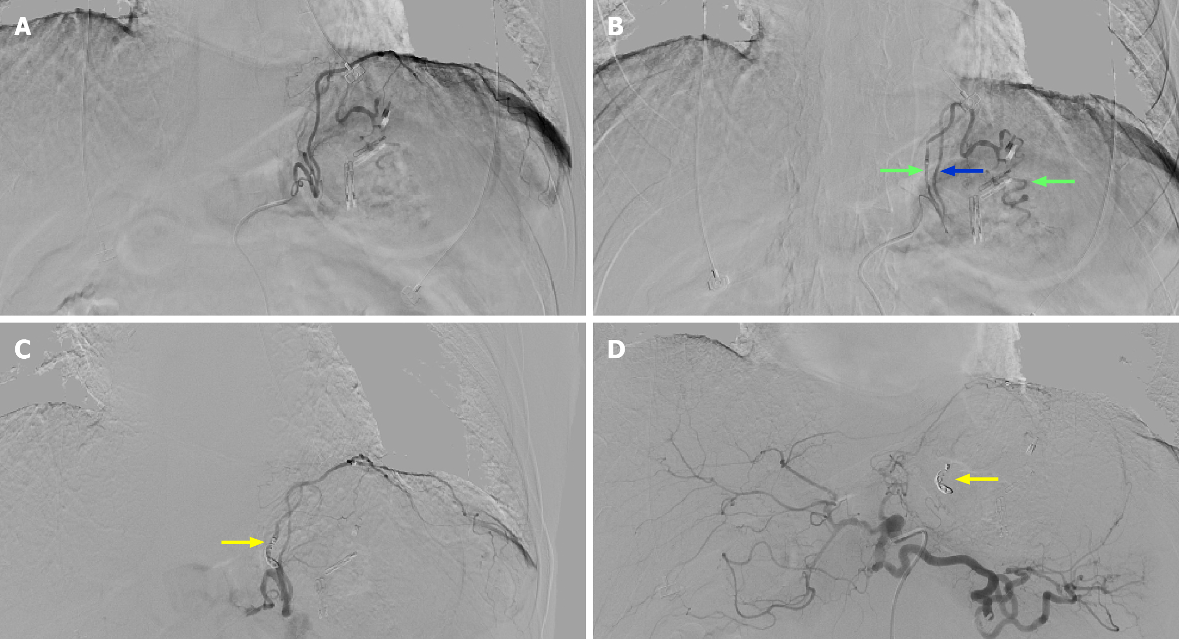
Figure 2 Abdominal angiography findings.
A: Two arteries from the celiac trunk supplying the spleen; B: The bleeding artery is one branch (indicated by green arrows) from the left inferior phrenic. Another branch (indicated by blue arrow) supplies the left diaphragmatic muscle; C: After transcatheter embolization with metallic coils (yellow arrow); D: Selective arteriography showed that the remaining dorsal pancreatic artery still supplies the spleen (yellow arrow).
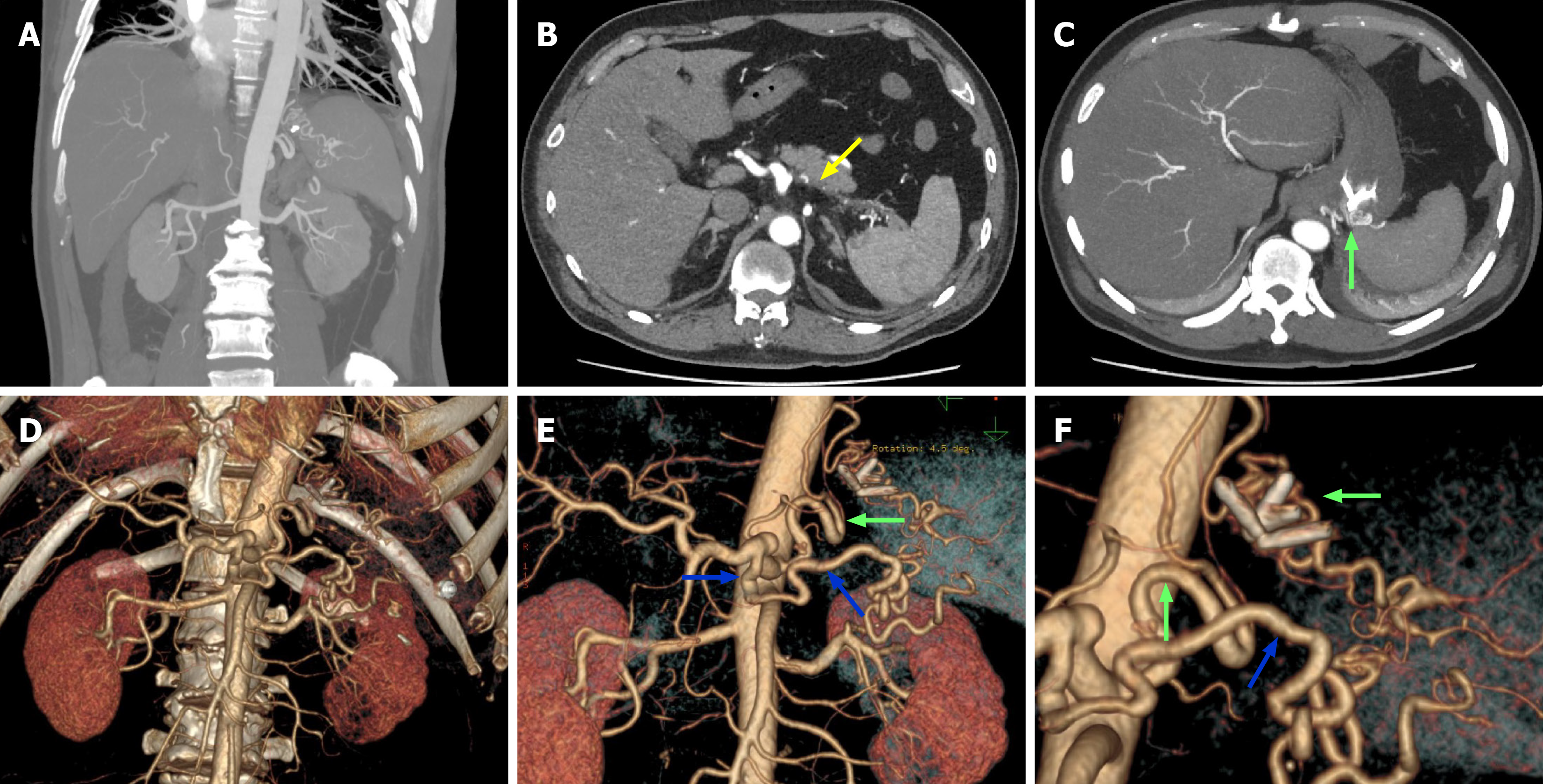
Figure 3 Portal vein computed tomography venography images.
A: Tortuous vessels supplying the spleen observed in the coronal view; B: The splenic artery occlusion indicated by yellow arrows; C: A tortuous artery running through the gastric wall indicated by green arrow; D: A three-dimensional computed tomography image shows the absence of the splenic artery; E and F: The tortuous artery running through the gastric wall (indicated by green arrows), which along with the enlarged dorsal pancreatic artery (indicated by blue arrows), supplies the spleen. The image shows the placement of clips on the gastric wall.
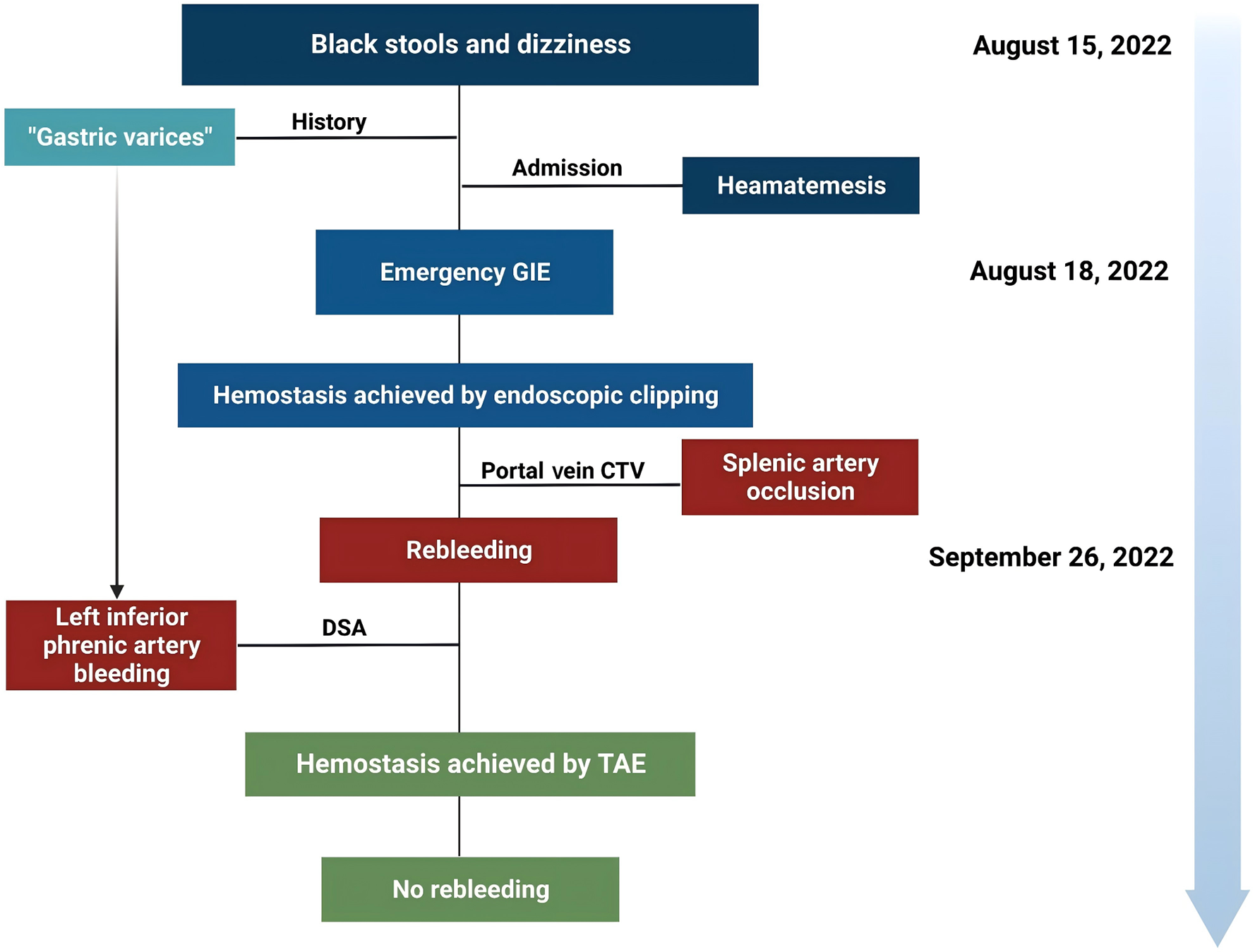
Figure 4 Medical timeline of the patient with acute upper gastrointestinal bleeding.
GIE: Gastrointestinal endoscopy; CTV: Computed tomography venography; DSA: Digital subtraction angiography; TAE: Transcatheter arterial embolization.
- Citation: Wang H, Tan YQ, Han P, Xu AH, Mu HL, Zhu Z, Ma L, Liu M, Xie HP. Left inferior phrenic arterial malformation mimicking gastric varices: A case report and review of literature. World J Gastrointest Surg 2024; 16(9): 3057-3064
- URL: https://www.wjgnet.com/1948-9366/full/v16/i9/3057.htm
- DOI: https://dx.doi.org/10.4240/wjgs.v16.i9.3057


