Copyright
©The Author(s) 2024.
World J Gastrointest Surg. Sep 27, 2024; 16(9): 3041-3047
Published online Sep 27, 2024. doi: 10.4240/wjgs.v16.i9.3041
Published online Sep 27, 2024. doi: 10.4240/wjgs.v16.i9.3041
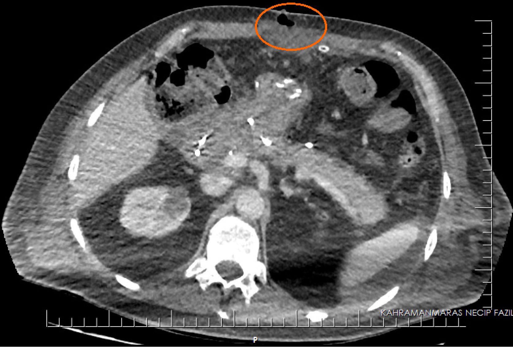
Figure 1 Computed tomography evaluation when subcutaneous collection was observed.
The marked area shows the collection.
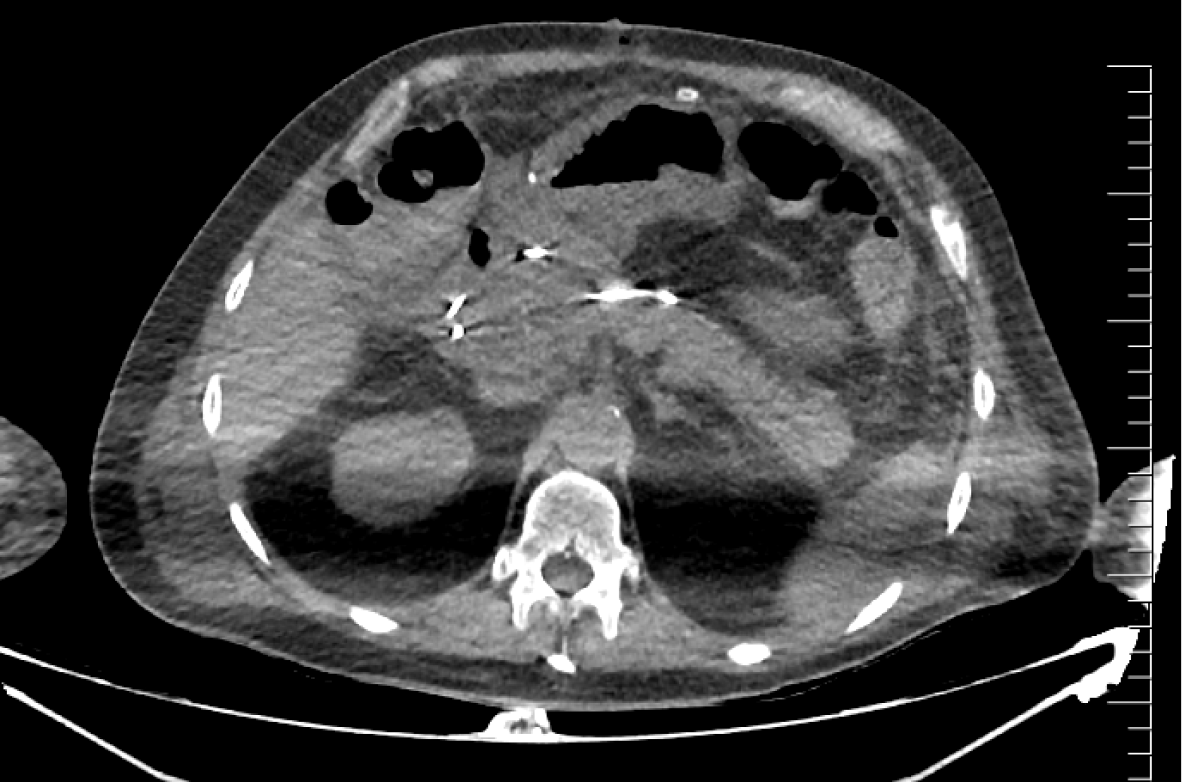
Figure 2 Control computed tomography imaging.
The image shows a displaced pancreatic stent.
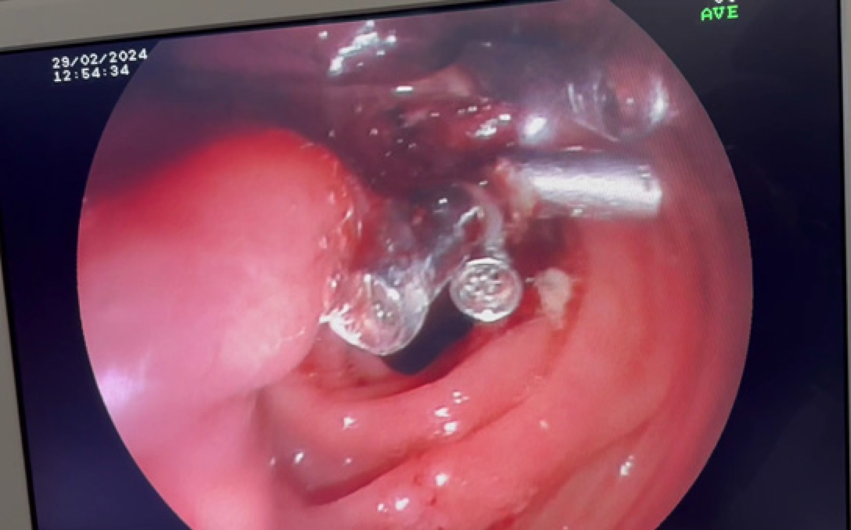
Figure 3 Endoscopic clipping.
Gastroenterostomy opening closure.
Figure 4 Preoperative imaging.
A: Preoperative magnetic resonance cholangiopancreatography examination. The marked area indicates the absence of contrast in the distal common bile duct; B: Preoperative upper abdominal magnetic resonance imaging examination. The marked area shows the pancreatic mass.
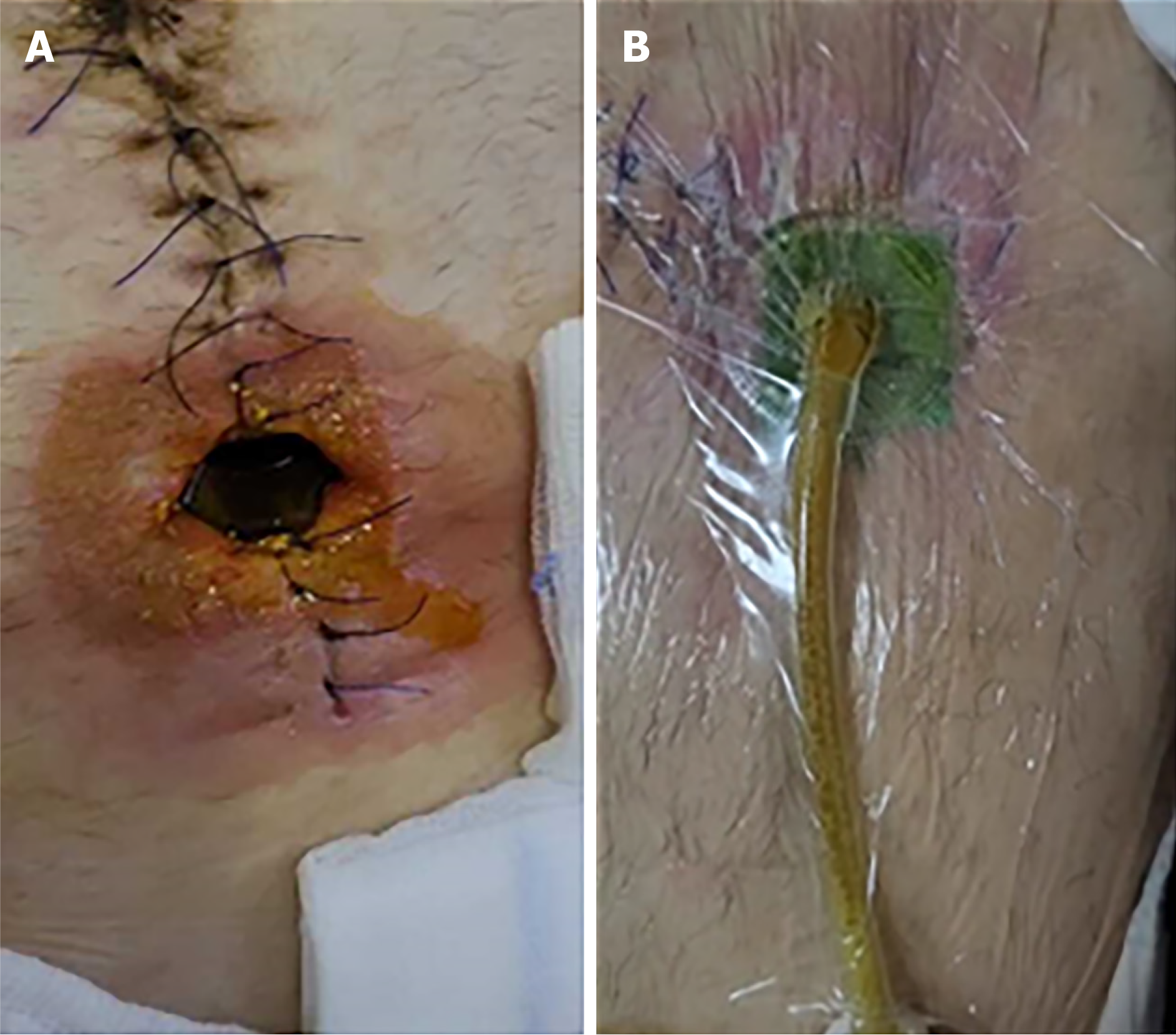
Figure 5 Physical examination findings.
A: Opening in the incision; B: Vacuum assisted closure therapy.
- Citation: Muhammedoğlu B, Ay OF. Endoscopic clipping of gastrojejunostomy leakage following Whipple procedure: A case report. World J Gastrointest Surg 2024; 16(9): 3041-3047
- URL: https://www.wjgnet.com/1948-9366/full/v16/i9/3041.htm
- DOI: https://dx.doi.org/10.4240/wjgs.v16.i9.3041


