Copyright
©The Author(s) 2024.
World J Gastrointest Surg. Sep 27, 2024; 16(9): 2853-2859
Published online Sep 27, 2024. doi: 10.4240/wjgs.v16.i9.2853
Published online Sep 27, 2024. doi: 10.4240/wjgs.v16.i9.2853
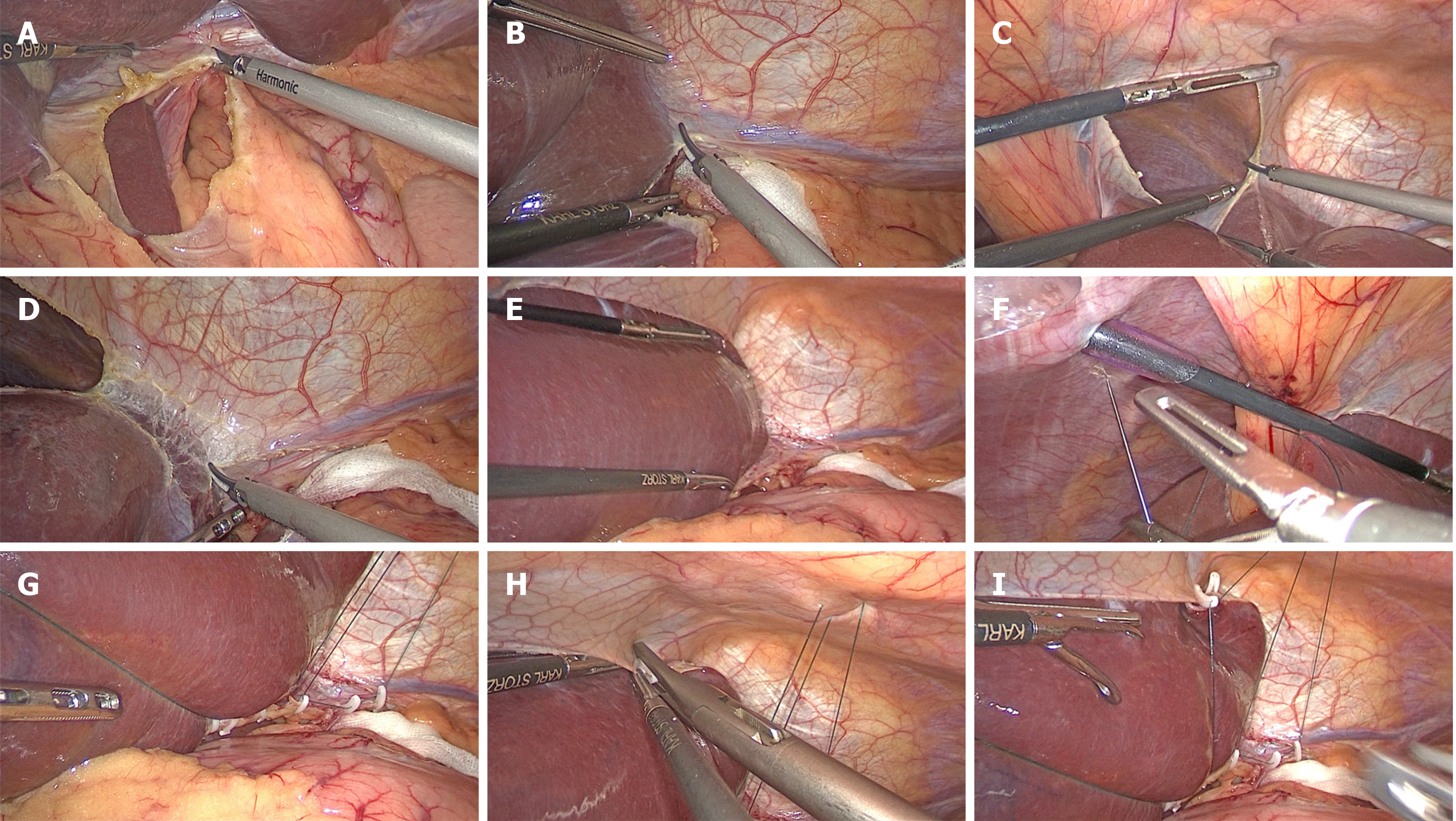
Figure 1 The procedures for the modified hepatic left lateral lobe inversion.
A: The hepatogastric ligament was opened; B: The left deltoid ligament was disconnected; C: The falciform ligament in the avascular area was dissected with an ultrasound knife or electric hook on the cephalic round ligament; D: The left coronary ligament was continuously dissected; E: After confirming the activity of the hepatic left lateral lobe, it was pushed through the dissected falciform ligament gap to the right superior hepatic space and inversed over; F: Subsequently, a 2-0 purse-string suture needle was inserted into the abdominal cavity from the left side of the falciform ligament, closely adhering to the xiphoid process caudally, followed by further insertion into the abdominal cavity close to the puncture point after perforation out the cavity, passing through the round ligament near the liver inside the cavity and then perforation out the cavity from the right side of the falciform ligament; G: The purse-string suture was fixed to the upper edge of the opened hepatogastric ligament using an absorbable clip below the liver; H: The inverse hepatic left lateral lobe was continued to be fixed to the upper edge of the dissected falciform ligament using an absorbable clip; I: The purse-string suture was tightened outside the body to obstruct the inverse hepatic left lateral lobe between the abdominal wall and the liver.
- Citation: Lin JA, Wu CY, Ye K. Modified hepatic left lateral lobe inversion in laparoscopic proximal gastrectomy: An analysis of 13 cases. World J Gastrointest Surg 2024; 16(9): 2853-2859
- URL: https://www.wjgnet.com/1948-9366/full/v16/i9/2853.htm
- DOI: https://dx.doi.org/10.4240/wjgs.v16.i9.2853


