Copyright
©The Author(s) 2024.
World J Gastrointest Surg. Aug 27, 2024; 16(8): 2592-2601
Published online Aug 27, 2024. doi: 10.4240/wjgs.v16.i8.2592
Published online Aug 27, 2024. doi: 10.4240/wjgs.v16.i8.2592
Figure 1 Methods of the new anti-mesenteric side-to-side delta-shaped stapled anastomosis and the conventional functional end-to-end anastomosis technique.
A: After a small anti-mesenteric incision was made, a linear stapler was inserted into the incision to perform the anastomosis; B: The conventional functional end-to-end anastomosis closed the open window transversely; C: The anti-mesenteric functional end-to-end delta-shaped stapled anastomosis technique was performed with a vertical closure of the open window using a linear stapler.
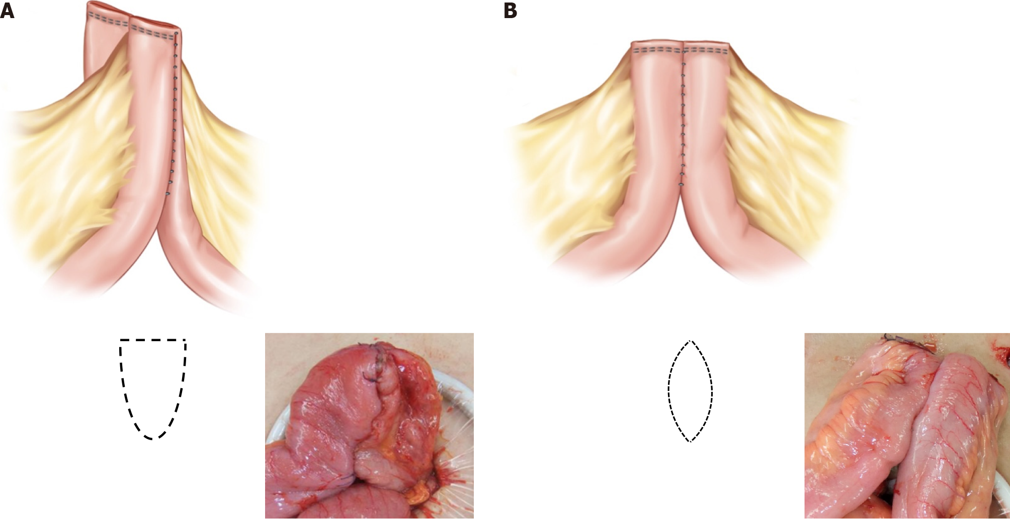
Figure 2 Comparison of intestinal lumen between the delta-shaped stapled anastomosis technique and the conventional functional end-to-end anastomosis technique.
A: When observing the anastomosis in the intestinal lumen, the anti-mesenteric functional end-to-end delta-shaped stapled anastomosis technique appears as a delta shape; B: An ovoid shape is formed after completion of the conventional functional end-to-end anastomosis.
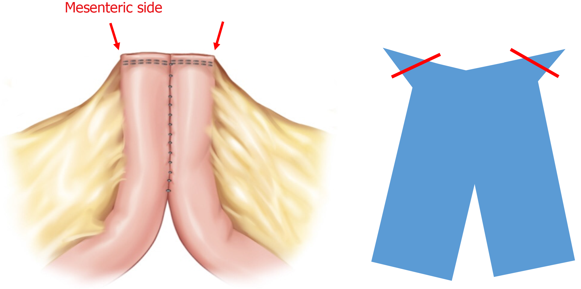
Figure 3 Exposure of the corner area after the conventional functional end-to-end anastomosis technique.
After performing conventional functional end-to-end anastomosis, the transverse end on the mesentery side protrudes, and the saccular or pouch-like part is formed.
- Citation: Lee JL, Yoon YS, Lee HG, Kim YI, Kim MH, Kim CW, Park IJ, Lim SB, Yu CS. New anti-mesenteric delta-shaped stapled anastomosis: Technical report with short-term postoperative outcomes in patients with Crohn’s disease. World J Gastrointest Surg 2024; 16(8): 2592-2601
- URL: https://www.wjgnet.com/1948-9366/full/v16/i8/2592.htm
- DOI: https://dx.doi.org/10.4240/wjgs.v16.i8.2592


