Copyright
©The Author(s) 2024.
World J Gastrointest Surg. Aug 27, 2024; 16(8): 2546-2554
Published online Aug 27, 2024. doi: 10.4240/wjgs.v16.i8.2546
Published online Aug 27, 2024. doi: 10.4240/wjgs.v16.i8.2546
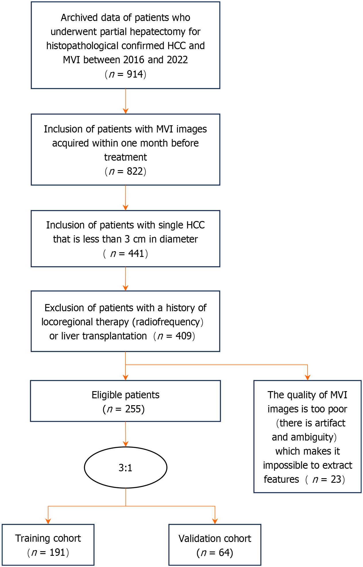
Figure 1 Flowchart of the search strategy used according to research requirements.
HCC: Hepatocellular carcinoma; MVI: Microvascular invasion.
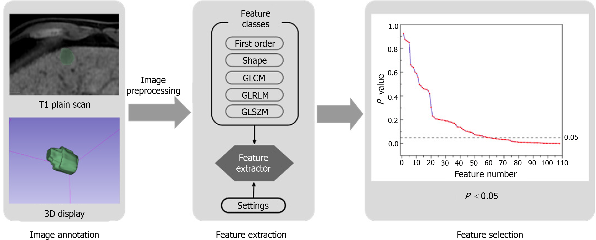
Figure 2 Tumor contours were labeled on each patient’s T1-weighted imaging plain image.
After preprocessing, image features were acquired by pyradiomics, and then the correlation between features and microvascular invasion. was analyzed. Features with P value < 0.05 were selected as input to the artificial neural network. GLCM: Grey level co-occurrence matrix; GLRLM: Grey level run length matrix; GLSZM: Gray level size zone matrix.
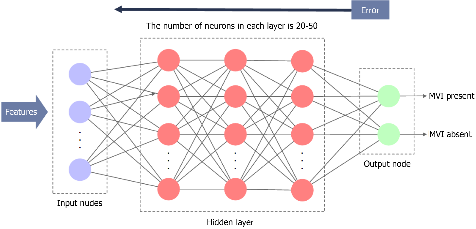
Figure 3 The basic structure of the artificial neural network constructed in this study.
MVI: Microvascular invasion.
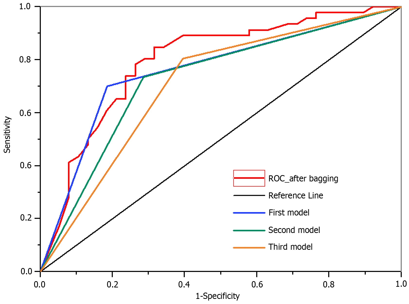
Figure 4 The results of the bagging strategy.
The red line represents achievement of area under the curve (AUC) of 0.79. The top three classifiers out of the 50 classifiers are represented by blue, green, and orange lines, corresponding to AUC values of 0.74, 0.73, and 0.71, respectively. ROC: Receiver operating characteristic.
- Citation: Xu JY, Yang YF, Huang ZY, Qian XY, Meng FH. Preoperative prediction of hepatocellular carcinoma microvascular invasion based on magnetic resonance imaging feature extraction artificial neural network. World J Gastrointest Surg 2024; 16(8): 2546-2554
- URL: https://www.wjgnet.com/1948-9366/full/v16/i8/2546.htm
- DOI: https://dx.doi.org/10.4240/wjgs.v16.i8.2546


