Copyright
©The Author(s) 2024.
World J Gastrointest Surg. Aug 27, 2024; 16(8): 2528-2537
Published online Aug 27, 2024. doi: 10.4240/wjgs.v16.i8.2528
Published online Aug 27, 2024. doi: 10.4240/wjgs.v16.i8.2528
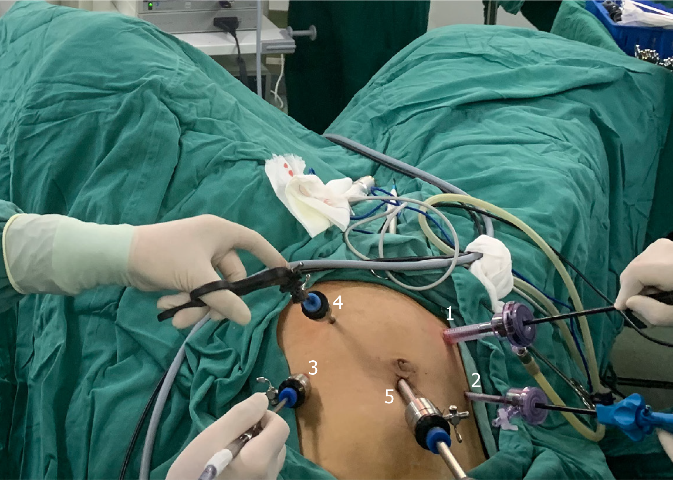
Figure 1 Layout of the trocars.
1: A 12 mm trocar is placed two transverse finger widths from the right anterior superior iliac spine as the main operation port for the surgeon; 2: A 5 mm trocar is placed at the right midclavicular line at the flat umbilicus as the auxiliary operation port for the surgeon; 3: A 5 mm trocar is placed at the left midclavicular line at the flat umbilicus as the main operation port for the first assistant; 4: A 5 mm trocar is placed at the left Mai’s point as an auxiliary operation port for the first assistant; 5: A 10 mm diameter trocar is placed 1 cm above the umbilicus as an observation port.
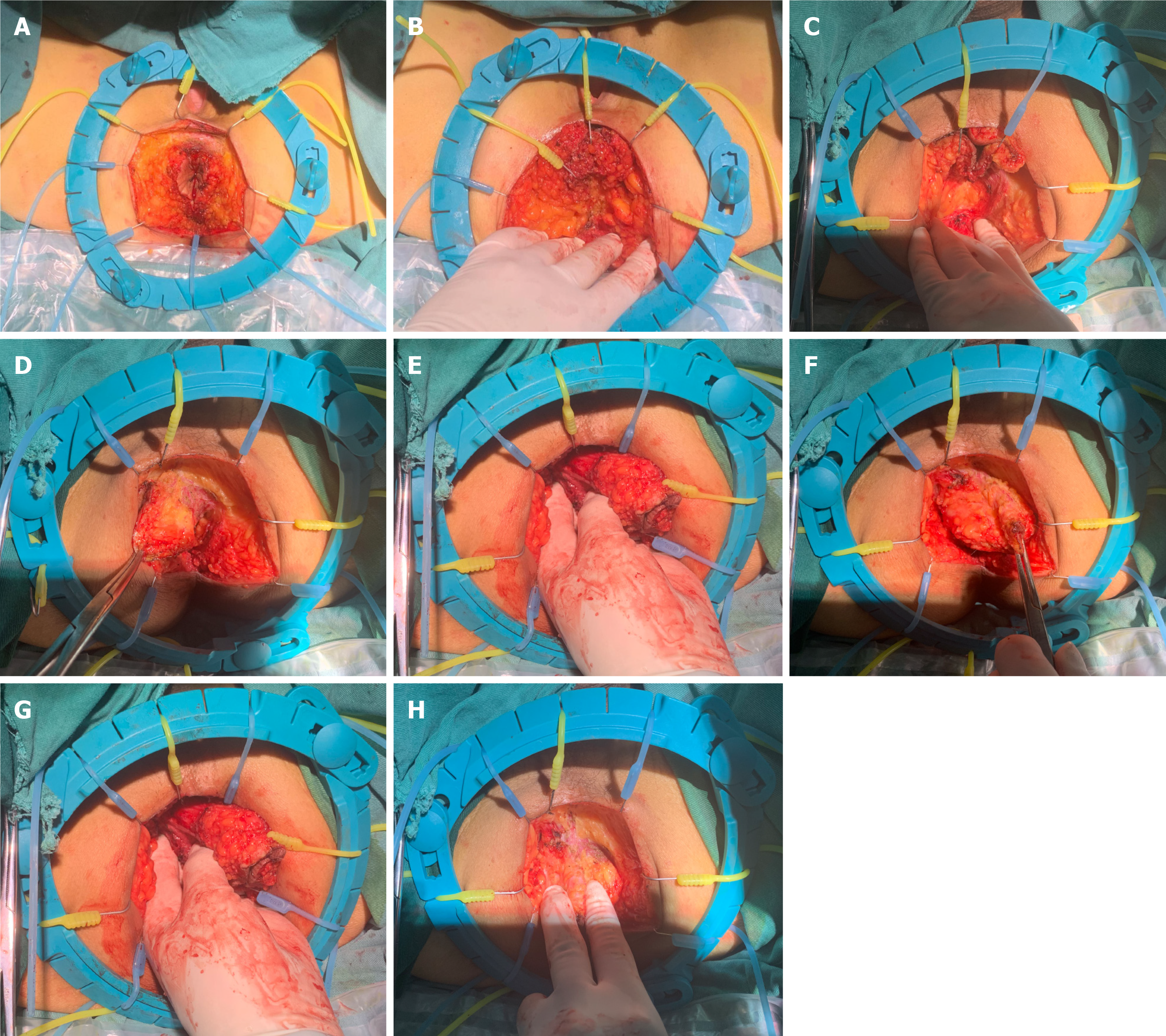
Figure 2 Application of the Lone-Star retractor method for perineal exposure.
A: After incision of the skin and subcutaneous tissue, a Lone-Star retractor is placed; B and C: Three small anterior hooks are used to pull the anal canal tissue toward 12:00; D and E: The left levator ani is dissected; F and G: The right levator ani is dissected; H: The anterior rectal wall tissue is detached.
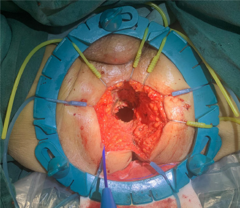
Figure 3 Display of the perineum after removal of the specimen.
- Citation: Ma J, Tang DB, Tang YQ, Wang DT, Jiang P, Zhang YM. Lone-Star retractor perineal exposure method for laparoscopic abdominal perineal resection of rectal cancer. World J Gastrointest Surg 2024; 16(8): 2528-2537
- URL: https://www.wjgnet.com/1948-9366/full/v16/i8/2528.htm
- DOI: https://dx.doi.org/10.4240/wjgs.v16.i8.2528


