Copyright
©The Author(s) 2024.
World J Gastrointest Surg. Aug 27, 2024; 16(8): 2474-2483
Published online Aug 27, 2024. doi: 10.4240/wjgs.v16.i8.2474
Published online Aug 27, 2024. doi: 10.4240/wjgs.v16.i8.2474
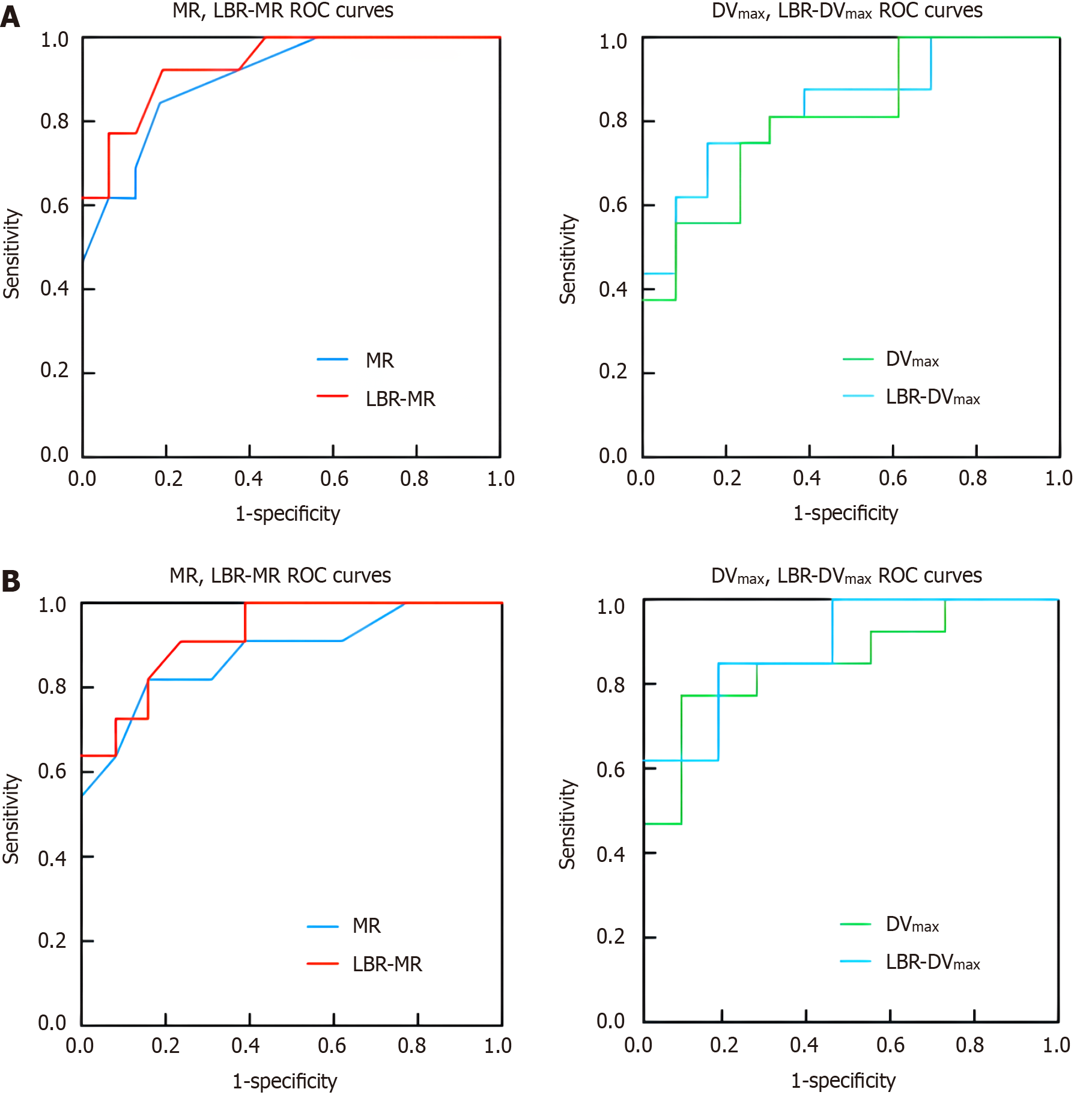
Figure 1 Receiver operating characteristic curve of dynamic positron emission tomography parameters in gastric cancer and esophageal cancer.
A: Receiver operating characteristic curve of dynamic positron emission tomography parameters in gastric cancer; B: Receiver operating characteristic curve of dynamic positron emission tomography parameters in esophageal cancer. MR: Metabolic rate; LBR: Lesion/background ratio; ROC: Receiver operating characteristic; DV: Volume distribution.
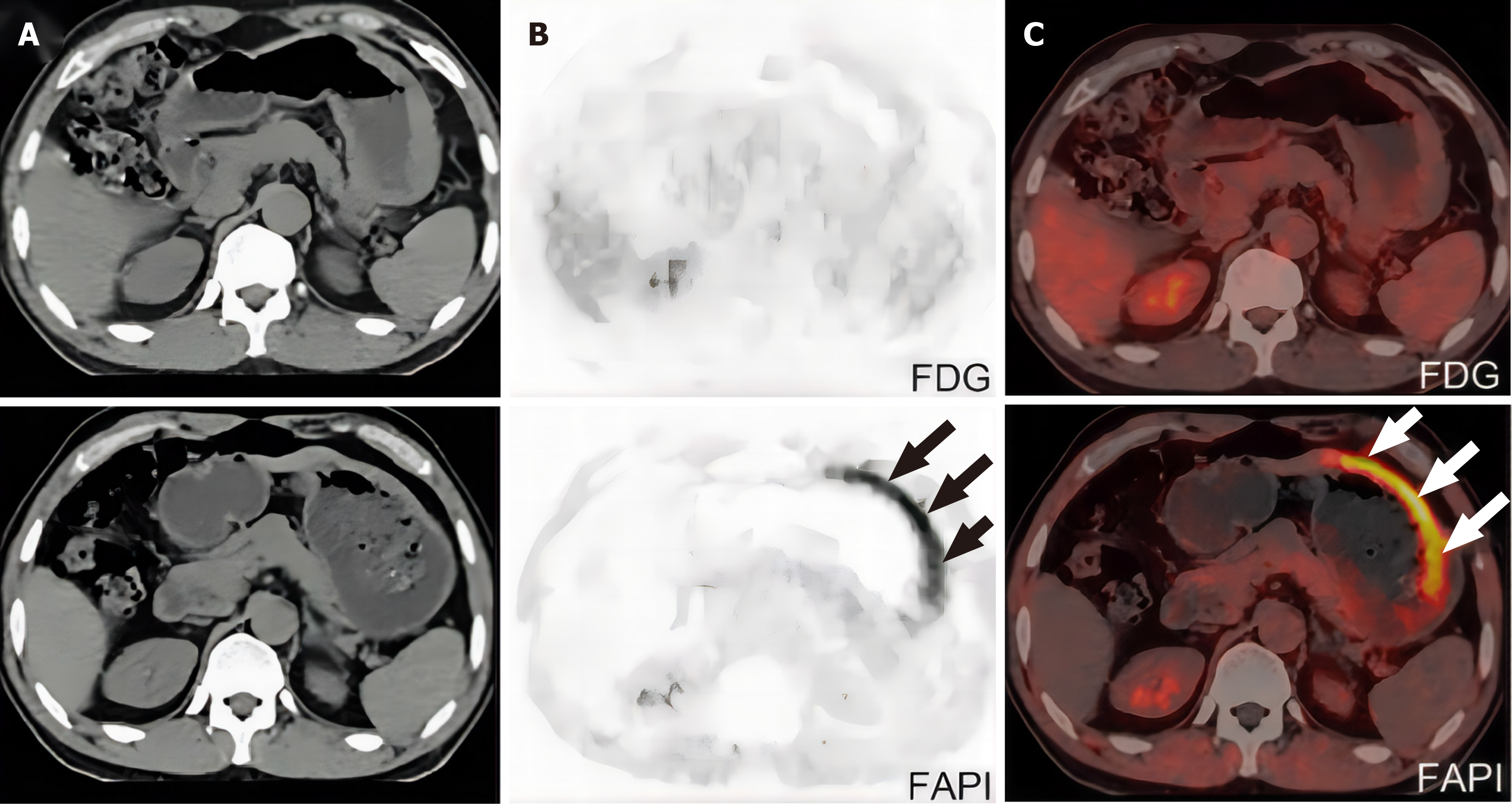
Figure 2 A patient with gastric signet-ring cell carcinoma underwent 18F-fluorodeoxyglucose-positron emission tomography/computed tomography for initial staging.
A: Computed tomography; B: 18F-fluorodeoxyglucose-FAPI-positron emission tomography/computed tomography; C: 68Ga-positron emission tomography/computed tomography. FDG: Fluorodeoxyglucose. Arrow: Tumor location.
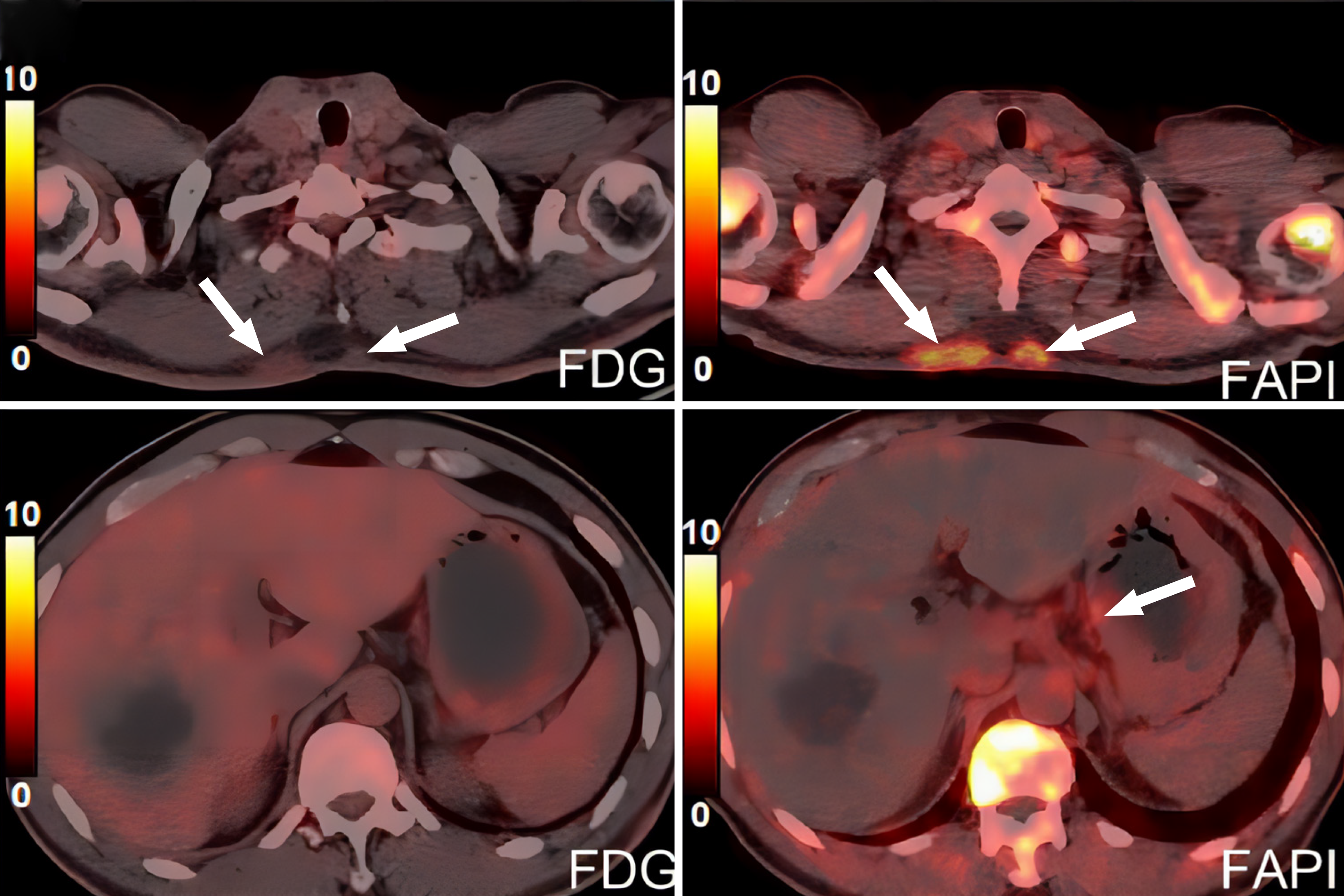
Figure 3 Subcutaneous and bone metastases of gastric cancer were identified by 18F-fluorodeoxyglucose positron emission tomography/computed tomography.
FDG: Fluorodeoxyglucose. Arrow: Tumor location.
- Citation: Ge DF, Ren H, Yang ZC, Zhao SX, Cheng ZT, Wu DD, Zhang B. Application of 18F-fluorodeoxyglucose positron emission tomography/computed tomography imaging in recurrent anastomotic tumors after surgery in digestive tract tumors. World J Gastrointest Surg 2024; 16(8): 2474-2483
- URL: https://www.wjgnet.com/1948-9366/full/v16/i8/2474.htm
- DOI: https://dx.doi.org/10.4240/wjgs.v16.i8.2474


