Copyright
©The Author(s) 2024.
World J Gastrointest Surg. Jul 27, 2024; 16(7): 2337-2342
Published online Jul 27, 2024. doi: 10.4240/wjgs.v16.i7.2337
Published online Jul 27, 2024. doi: 10.4240/wjgs.v16.i7.2337
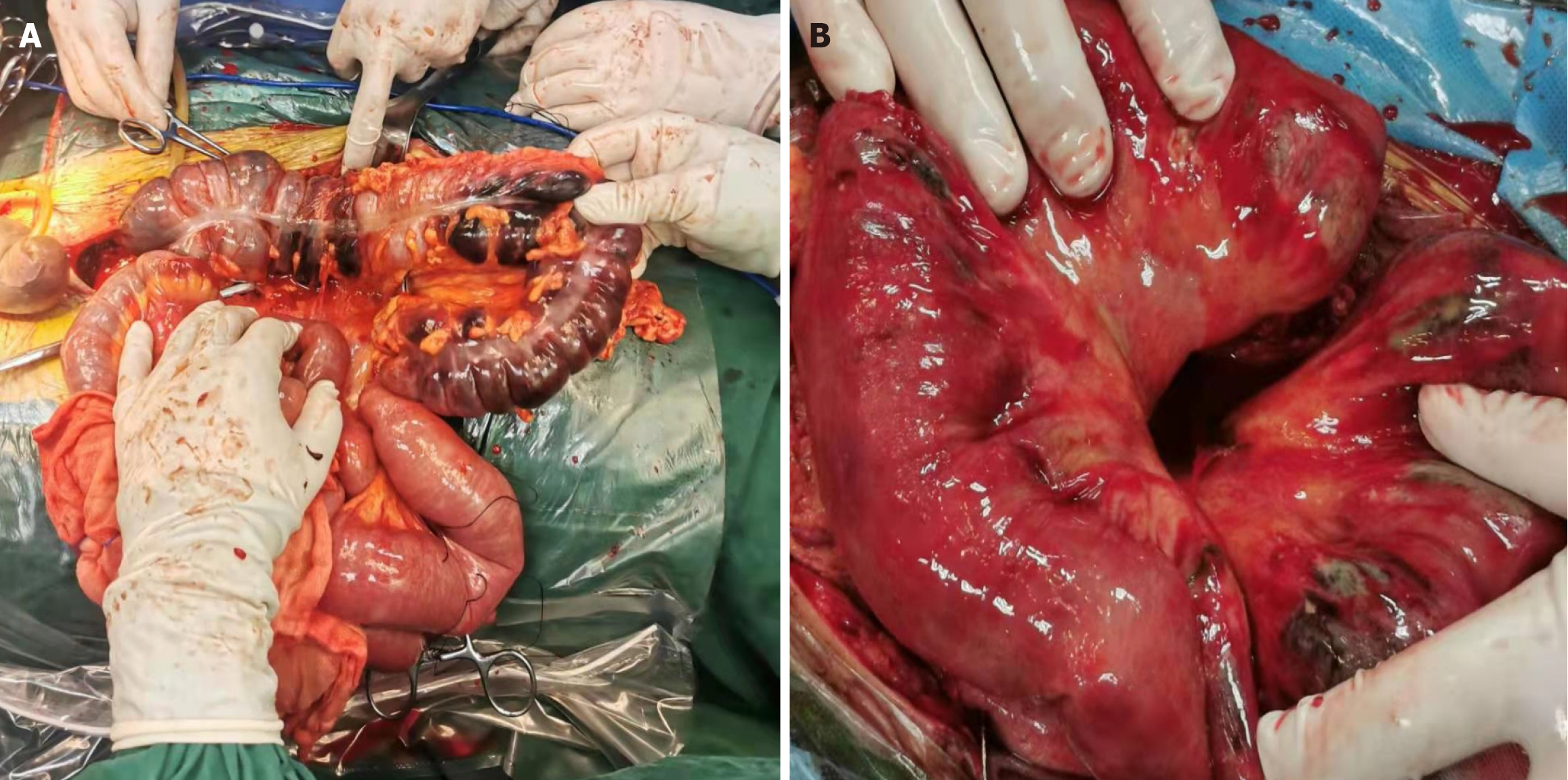
Figure 1 Intestinal necrosis was observed during the surgical procedure.
A: Necrosis of the colon was observed during the first exploratory laparotomy; B: Ischemic necrosis of the jejunum was observed during the second exploratory laparotomy.
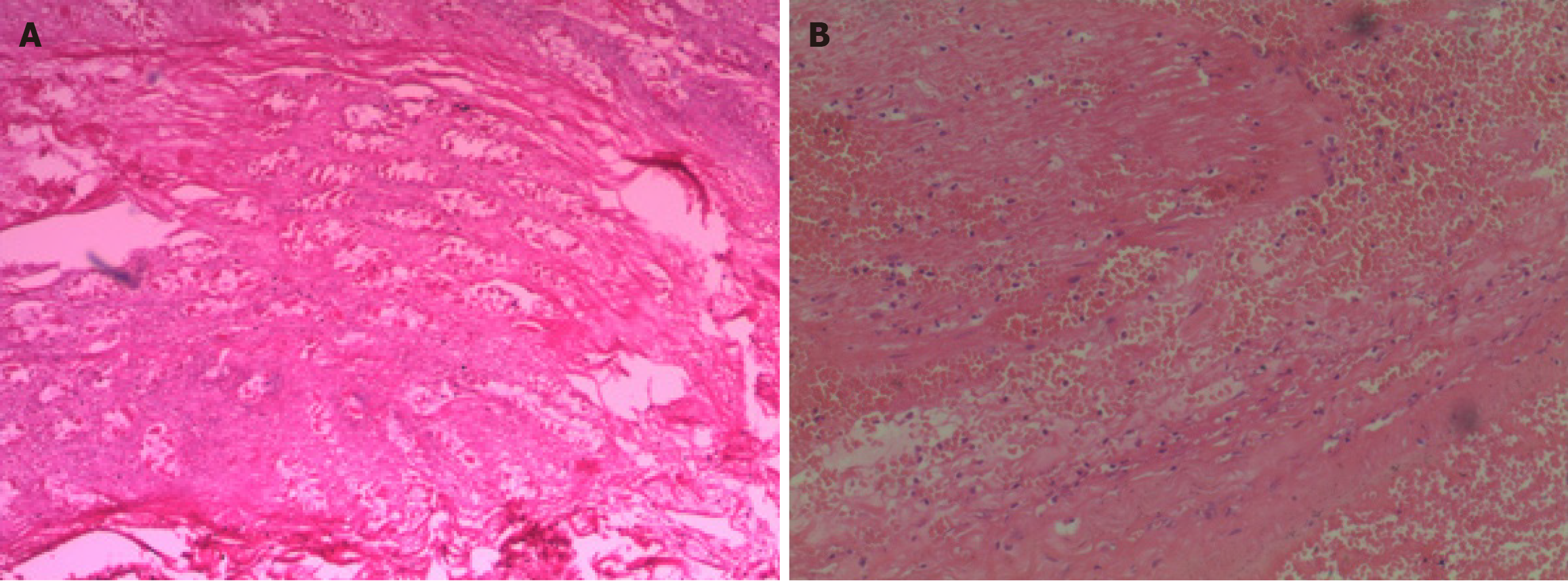
Figure 2 Histopathological examination revealed the bowel necrosis.
A: Histopathological analysis suggesting extensive bleeding and necrosis of the colon (× 100); B: Histopathological analysis suggesting bleeding from the small bowel rupture with acute purulent inflammation (× 100).
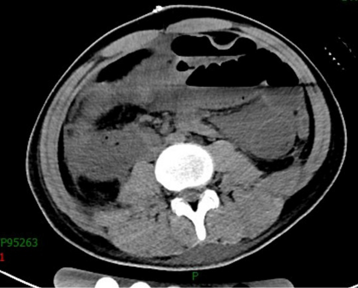
Figure 3
The abdomen computed tomography scan reveals multiple intestinal dilation, fluid and gas, and edema of the intestinal wall.
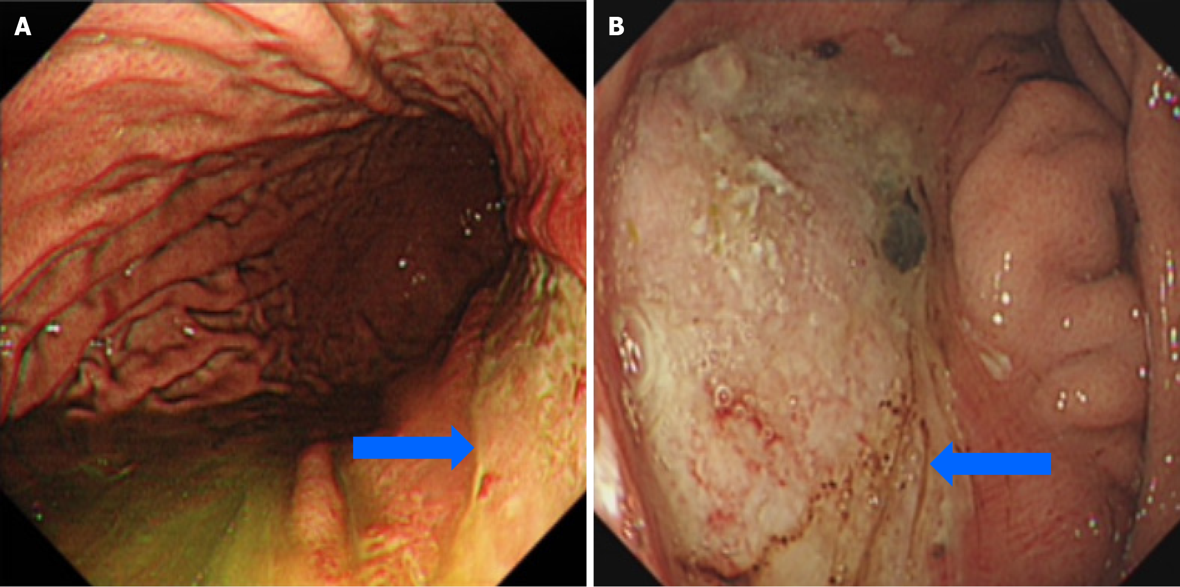
Figure 4 Gastroduodenoscopic images of gastric ulcer before and after the treatment.
A: Gastroduodenoscopic image showing a large band ulcer in the gastric lesser curvature; B: Gastroduodenoscopic image showing that the size of the ulcer was significantly reduced after treatment.
- Citation: Wang P, Wang TG, Yu AY. Sequential bowel necrosis and large gastric ulcer in a patient with a ruptured femoral artery: A case report. World J Gastrointest Surg 2024; 16(7): 2337-2342
- URL: https://www.wjgnet.com/1948-9366/full/v16/i7/2337.htm
- DOI: https://dx.doi.org/10.4240/wjgs.v16.i7.2337


