Copyright
©The Author(s) 2024.
World J Gastrointest Surg. Jul 27, 2024; 16(7): 1986-2002
Published online Jul 27, 2024. doi: 10.4240/wjgs.v16.i7.1986
Published online Jul 27, 2024. doi: 10.4240/wjgs.v16.i7.1986
Figure 1 Transabdominal ultrasonography.
A: Pseudocyst of the body-tail of the pancreas in the setting of chronic pancreatitis (indicated by an arrow); B: Pseudocyst of the head of the pancreas (indicated by an arrow).
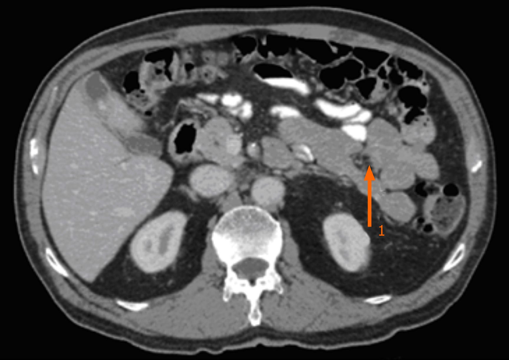
Figure 2
Computed tomography scan image showing pseudocyst at the tail of the pancreas (indicated by an arrow).
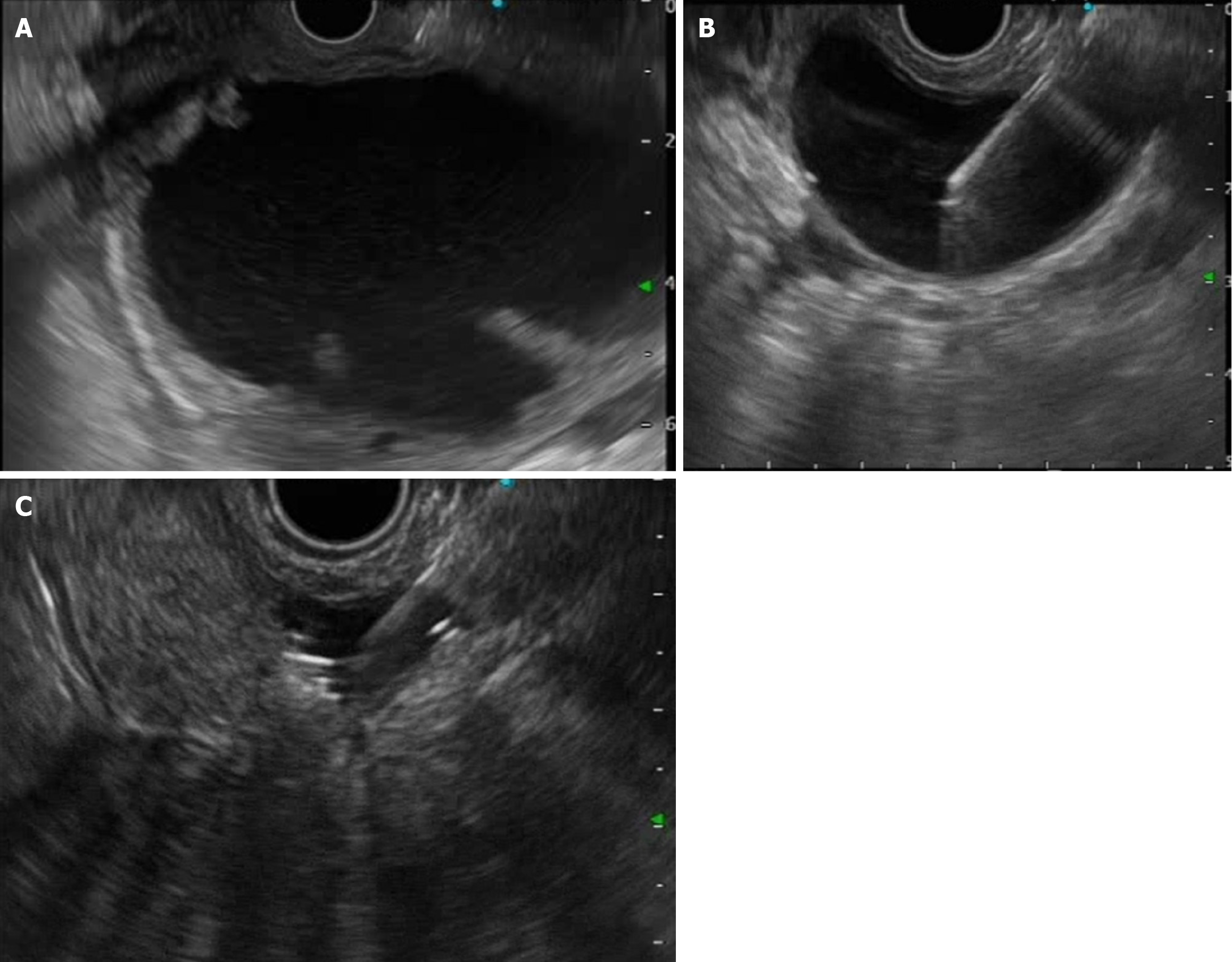
Figure 3 Endoscopic ultrasound utility in diagnosis and management of pancreas pseudocyst.
A: Appearance of pseudocyst on endoscopic ultrasound; B: Endoscopic ultrasound-guided fine-needle aspiration of pseudocyst; C: Decrease in size of pseudocyst after aspiration of cyst fluid.
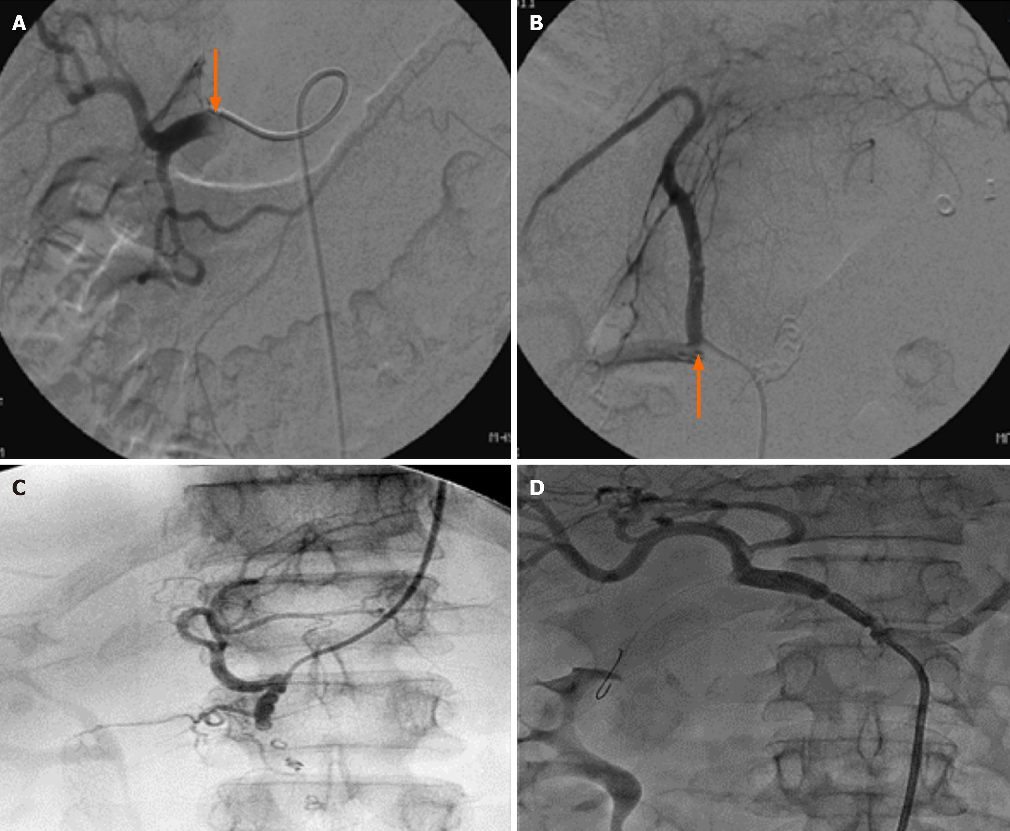
Figure 4 Angiogram.
A: Bleeding into the pseudocyst cavity from gastroduodenal artery (indicated by an arrow); B: Bleeding into the pseudocyst cavity (“cut off” of the left gastric artery, indicated by an arrow); C: Embolization was performed distal and proximal to the erosion zone; D: A stent graft was placed in the common hepatic artery.
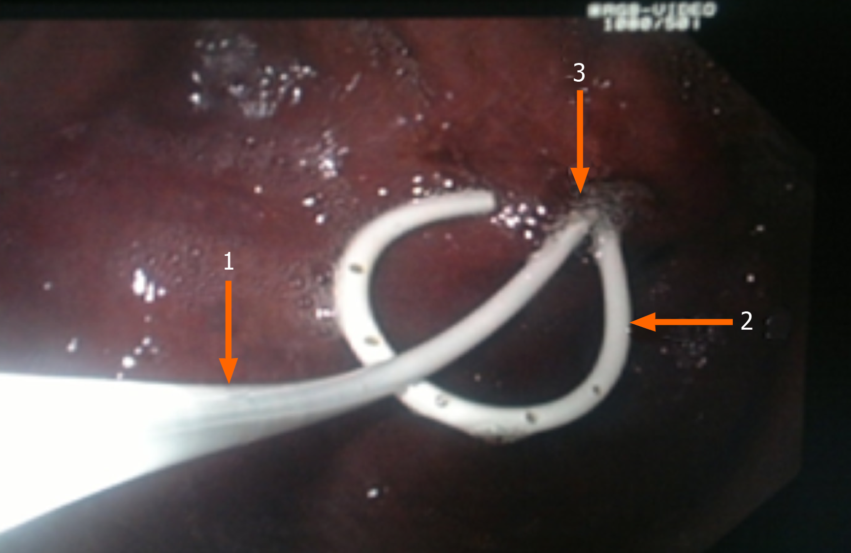
Figure 5 Transmural cystogastroanastomosis with external-internal drainage.
Arrow 1 shows cystonasal drainage, arrow 2 shows internal stent, and arrow 3 shows cystogastroanastomosis.
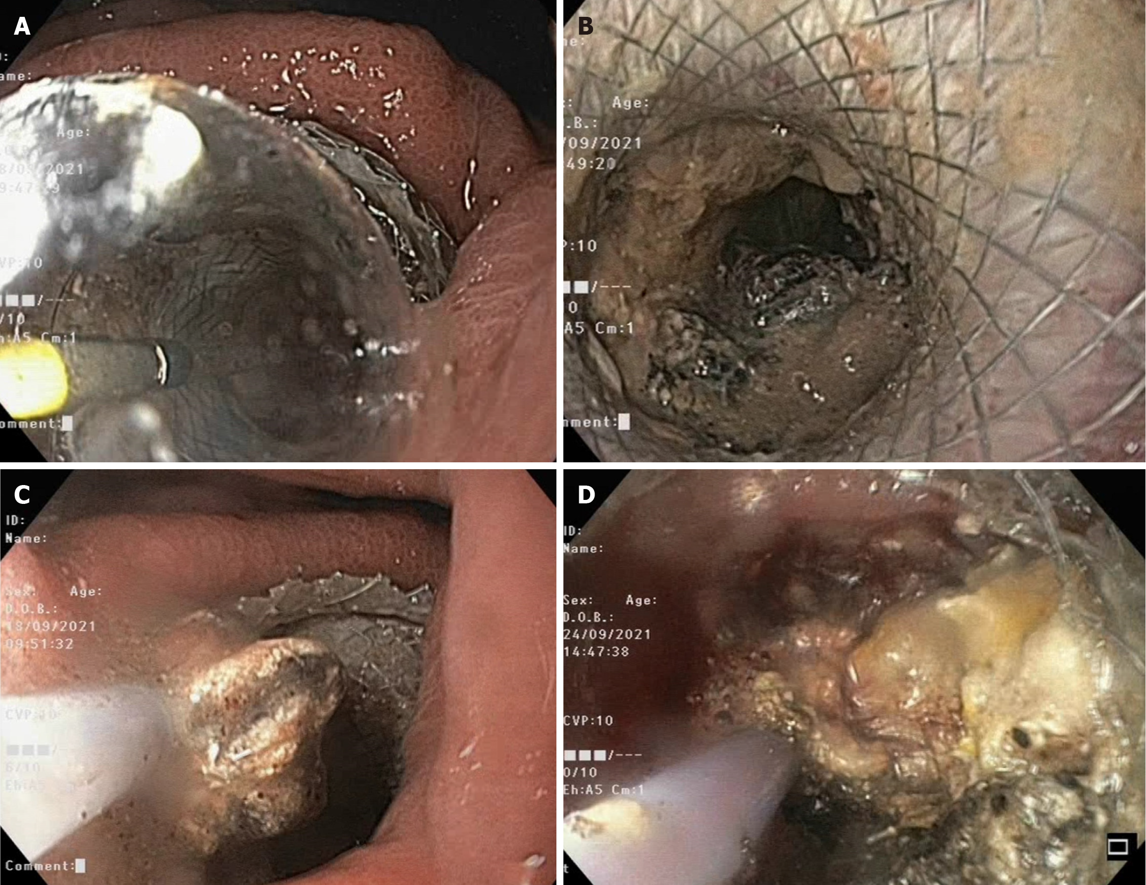
Figure 6 Endoscopic images.
A: Balloon dilatation of a lumen-apposing metal stent; B: The view inside the walled-off necrosis cavity after cystgastrostomy; C: Direct endoscopic necrosectomy with removal of debris using an endoscopic snare device; D: Bleeding within walled-off necrosis cavity during direct endoscopic necrosectomy.
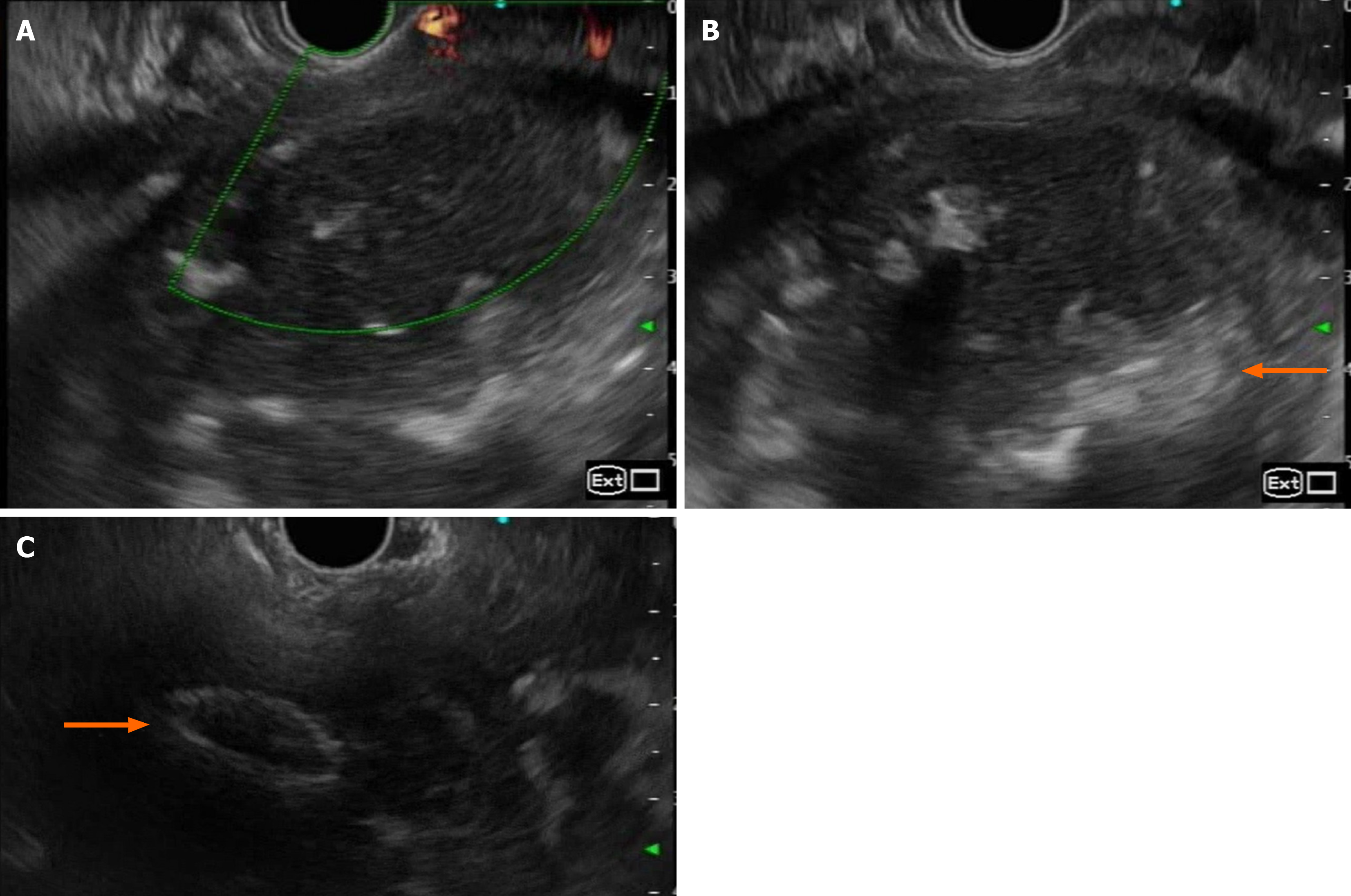
Figure 7 Endoscopic ultrasound images.
A: Absence of Doppler signal within walled-off necrosis cavity, ruling out presence of vessels or pseudoaneurysm; B: Solid debris (labelled with arrow) seen within walled-off necrosis cavity; C: Post deployment of lumen-apposing metal stent (labelled with arrow).
Figure 8 Percutaneous drain and dye study in pancreas pseudocyst management.
A: Percutaneous drainage of an infected pancreatic pseudocyst via a pigtail drain; B: Lavage of the cavity of the infected pseudocyst; C: Cystography in which the cavity of the pseudocyst was visualized, and the pigtail drainage was established.
- Citation: Koo JG, Liau MYQ, Kryvoruchko IA, Habeeb TA, Chia C, Shelat VG. Pancreatic pseudocyst: The past, the present, and the future. World J Gastrointest Surg 2024; 16(7): 1986-2002
- URL: https://www.wjgnet.com/1948-9366/full/v16/i7/1986.htm
- DOI: https://dx.doi.org/10.4240/wjgs.v16.i7.1986


