Copyright
©The Author(s) 2024.
World J Gastrointest Surg. Jun 27, 2024; 16(6): 1933-1938
Published online Jun 27, 2024. doi: 10.4240/wjgs.v16.i6.1933
Published online Jun 27, 2024. doi: 10.4240/wjgs.v16.i6.1933
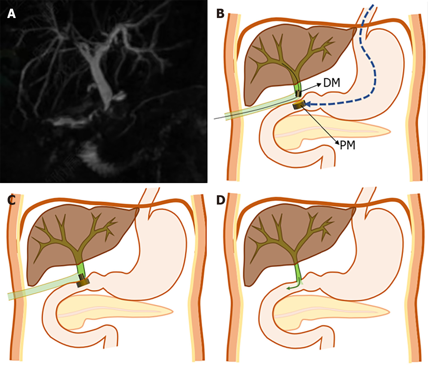
Figure 1 The surgical planning of abdominal sinus-duodenal magnetic compression anastomosis.
A: Magnetic resonance cholangio
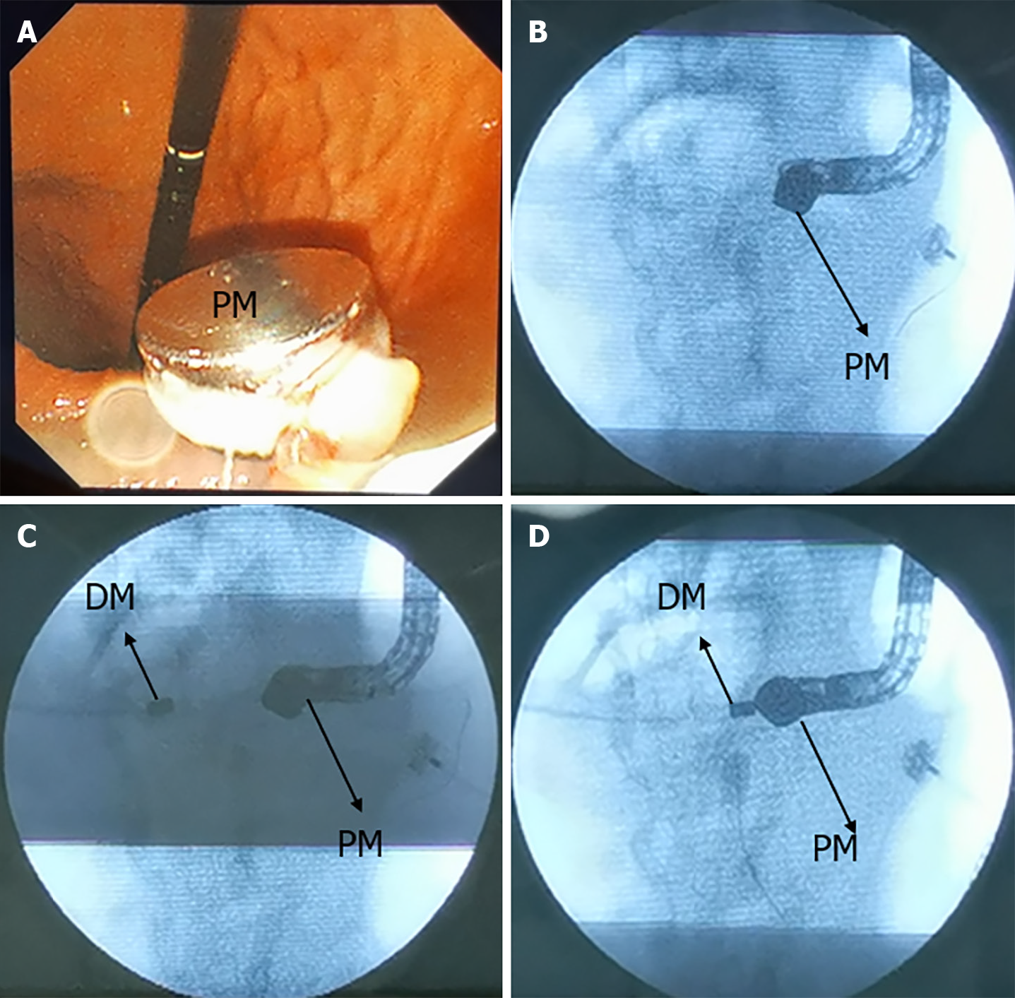
Figure 2 Endoscopic procedure.
A: The parent magnet (PM) enters the duodenum; B: The PM is shown in X-ray; C: The daughter magnet (DM) is inserted through the abdominal sinus; D: X-ray shows that the DM is attracted to the PM. DM: Daughter magnet; PM: Parent magnet.
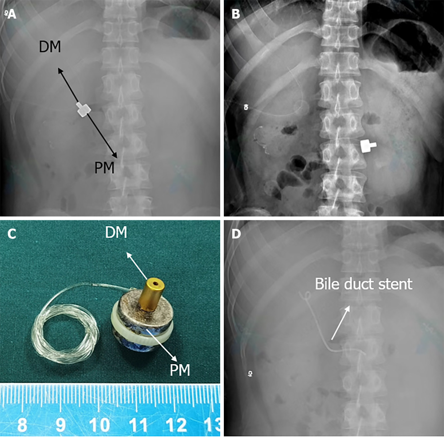
Figure 3 Postoperative abdominal imaging data.
A: The state of the magnets one day after the operation; B: The state of the magnets three days after the operation; C: The magnets expelled from the body; D: Biliary stent has been placed at the abdominal sinus-duodenal magnetic anastomosis. DM: Daughter magnet; PM: Parent magnet.
- Citation: Zhang MM, Tao J, Sha HC, Li Y, Song XG, Muensterer OJ, Dong FF, Zhang L, Lyu Y, Yan XP. Magnetic compression anastomosis to restore biliary tract continuity after obstruction following major abdominal trauma: A case report. World J Gastrointest Surg 2024; 16(6): 1933-1938
- URL: https://www.wjgnet.com/1948-9366/full/v16/i6/1933.htm
- DOI: https://dx.doi.org/10.4240/wjgs.v16.i6.1933


