Copyright
©The Author(s) 2024.
World J Gastrointest Surg. Jun 27, 2024; 16(6): 1910-1917
Published online Jun 27, 2024. doi: 10.4240/wjgs.v16.i6.1910
Published online Jun 27, 2024. doi: 10.4240/wjgs.v16.i6.1910
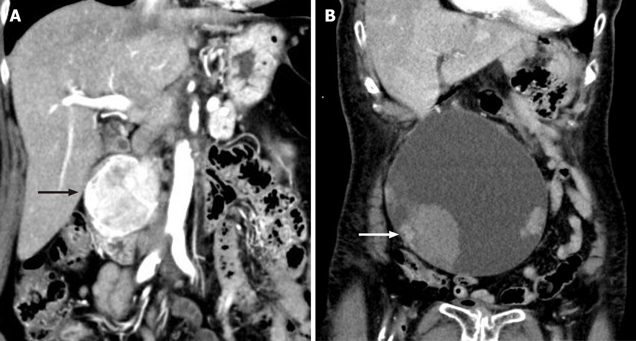
Figure 1 Imaging of the duodenal tumor and pancreatic tumor.
A: Imaging of the duodenal tumor and pancreatic tumor. Contrast-enhanced computed tomography showed a well-defined, enhancing masse, sized 5.0 cm × 6.0 cm, with heterogeneous density at the second portion of the duodenum (arrow); B: Imaging of the pancreatic tumor. Contrast-enhanced computed tomography showed a huge cystic mass measuring 17 cm, with some solid components and adjacent hematoma (arrow).
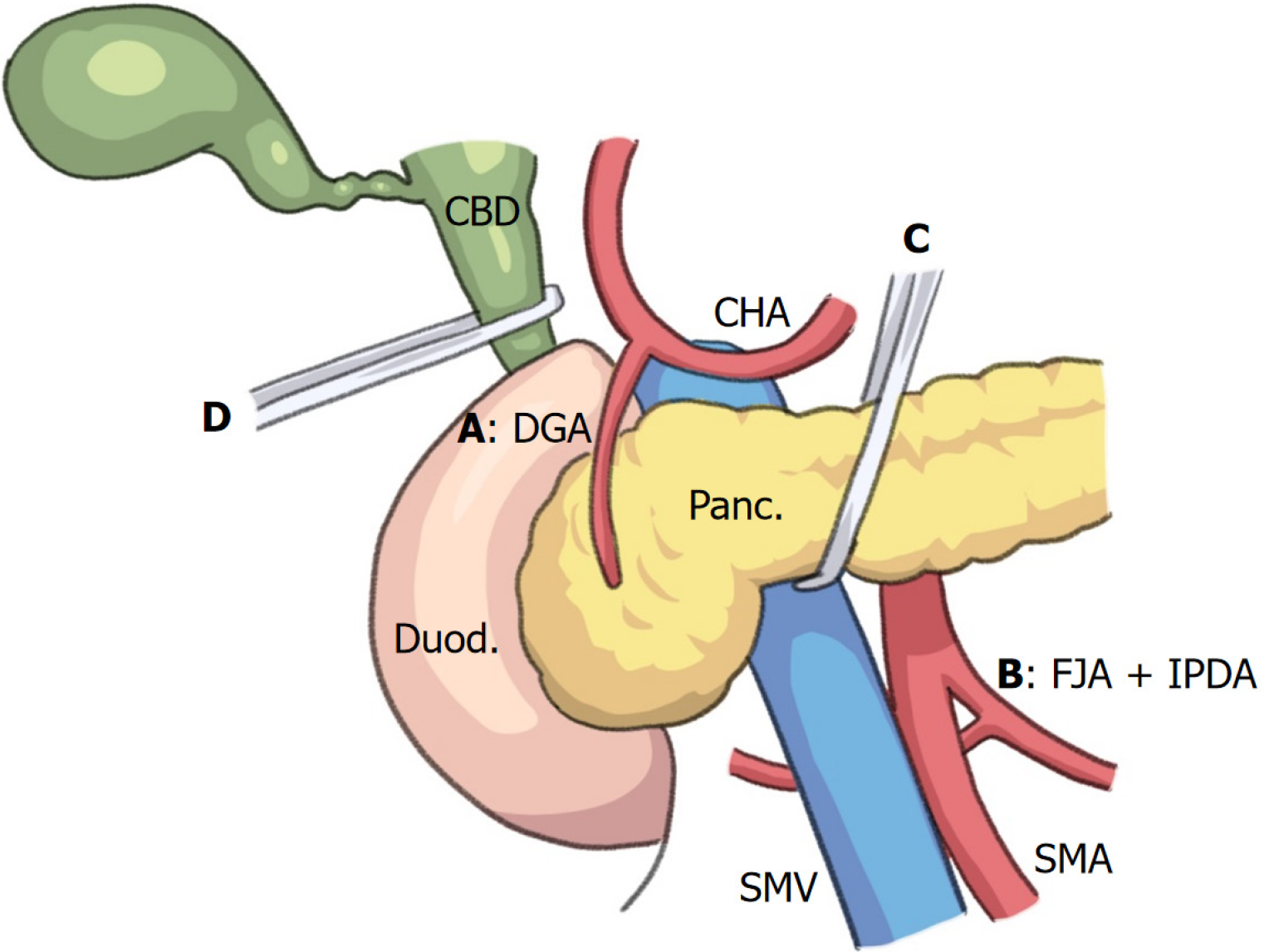
Figure 2 Illustration of no-touch isolation technique in emergency pancreaticoduodenectomy.
All arteries (A-D) that supply the pancreatic head region are isolated before manipulation of the pancreatic head and duodenum in the early phase of resection during emergency pancreaticoduodenectomy. A: The gastroduodenal artery; B: First jejunal artery, inferior pancreaticoduodenal artery; C: Intrapancreatic arterial arcade, including the inferior pancreatic artery. D: The para-biliary arterial plexus, including the 3 or 9 o’clock arteries, anastomoses to the posterior superior pancreaticoduodenal artery. Panc: Pancreas; Duod: Duodenum; SMV: Superior mesenteric vein; SMA: Superior mesenteric artery; CHA: Common hepatic artery; CBD: Common bile duct; GDA: Gastroduodenal artery; IPDA: Inferior pancreaticoduodenal artery; FJA: First jejunal artery.
- Citation: Cho A, Katagiri S, Ota M, Onizawa S, Higuchi R, Sugishita T, Niwa Y, Ishita T, Mouri T, Kato A, Iwata M. No-touch isolation technique in emergency pancreaticoduodenectomy for neoplastic hemorrhage: Two case reports and review of literature. World J Gastrointest Surg 2024; 16(6): 1910-1917
- URL: https://www.wjgnet.com/1948-9366/full/v16/i6/1910.htm
- DOI: https://dx.doi.org/10.4240/wjgs.v16.i6.1910


