Copyright
©The Author(s) 2024.
World J Gastrointest Surg. Jun 27, 2024; 16(6): 1548-1557
Published online Jun 27, 2024. doi: 10.4240/wjgs.v16.i6.1548
Published online Jun 27, 2024. doi: 10.4240/wjgs.v16.i6.1548
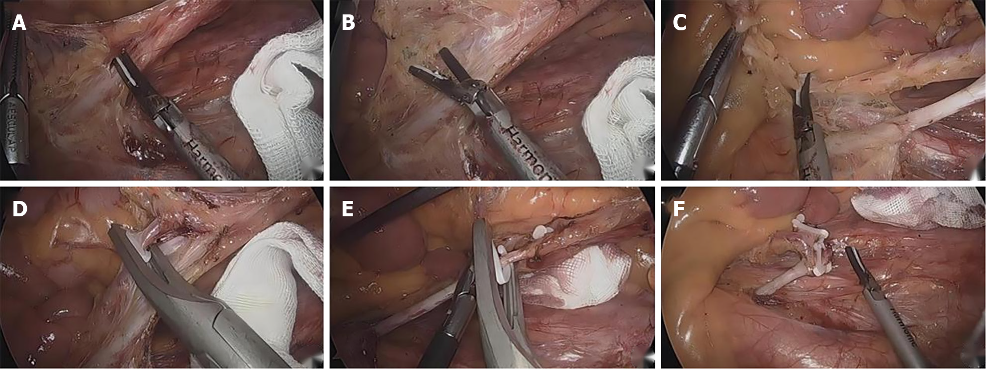
Figure 1 The left colic artery was preserved.
A: Isolation of the main naked inferior mesenteric artery; B: Isolatedisolation of the naked left colic artery; C: Clearance of the lymph nodes in group 253; D: Ligation and dissection of the sigmoid artery; E: Ligation of the severed superior rectal artery; F: Operation.
Figure 2 Submesenteric artery classification model.
A: Type I; B: Type II; C: Type III; D: Type IV. IMA: Inferior mesenteric artery; LCA: Left colic artery; SA: Sigmoid artery.
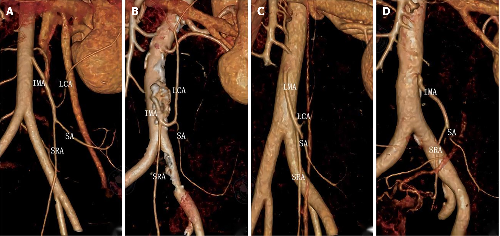
Figure 3 3D reconstruction of submesenteric artery typing.
A: Type I; B: Type II; C: Type III; D: Type IV. IMA: Inferior mesenteric artery; LCA: Left colic artery; SA: Sigmoid artery; SRA: Superior rectal artery.
- Citation: Wang Y, Liu ZS, Wang ZB, Liu S, Sun FB. Efficacy of laparoscopic low anterior resection for colorectal cancer patients with 3D-vascular reconstruction for left coronary artery preservation. World J Gastrointest Surg 2024; 16(6): 1548-1557
- URL: https://www.wjgnet.com/1948-9366/full/v16/i6/1548.htm
- DOI: https://dx.doi.org/10.4240/wjgs.v16.i6.1548


