Copyright
©The Author(s) 2024.
World J Gastrointest Surg. May 27, 2024; 16(5): 1474-1481
Published online May 27, 2024. doi: 10.4240/wjgs.v16.i5.1474
Published online May 27, 2024. doi: 10.4240/wjgs.v16.i5.1474
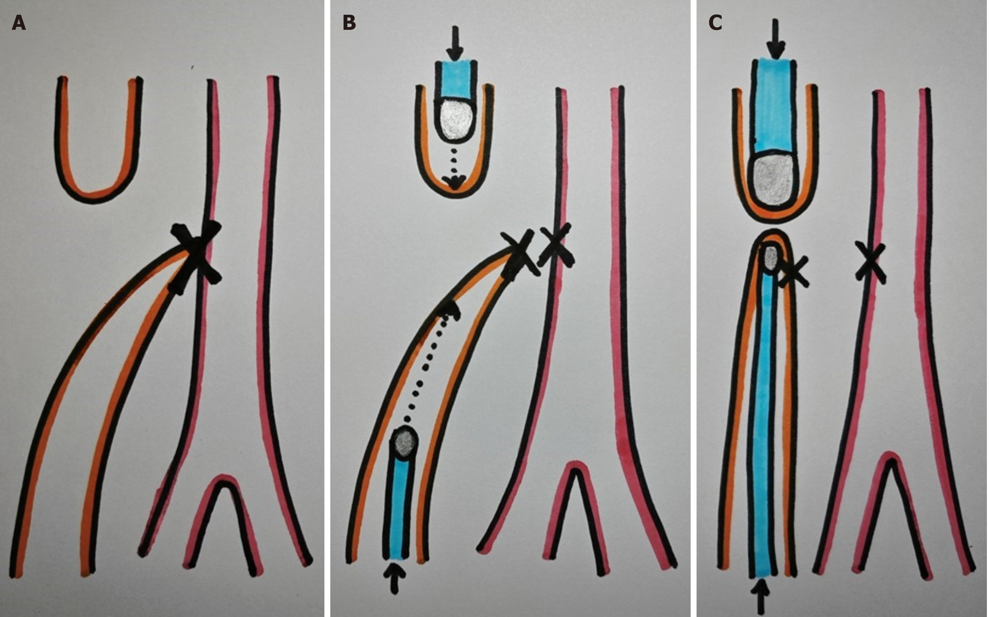
Figure 1 Alternative esophageal end-to-side anastomosis.
A: Esophageal atresia Gross Type C: The X indicates the area of ligation and section of the distal tracheoesophageal fistula; B: Magnets Advancement: Magnets are carefully advanced through each esophageal pouch using catheters to push them; C: Avoiding Suture Interposition: To prevent suture interposition in the distal esophageal pouch, the distal magnet is positioned laterally to the stitches, creating an end-to-side anastomosis. The X denotes the area of suture in the trachea and distal esophagus.
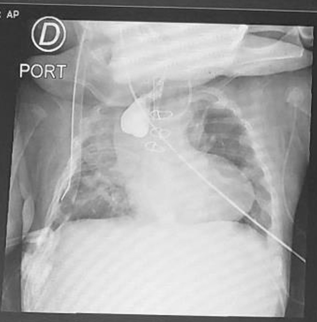
Figure 2 Esophagogram showing complete obliteration of the anastomosis due to severe esophageal stricture.
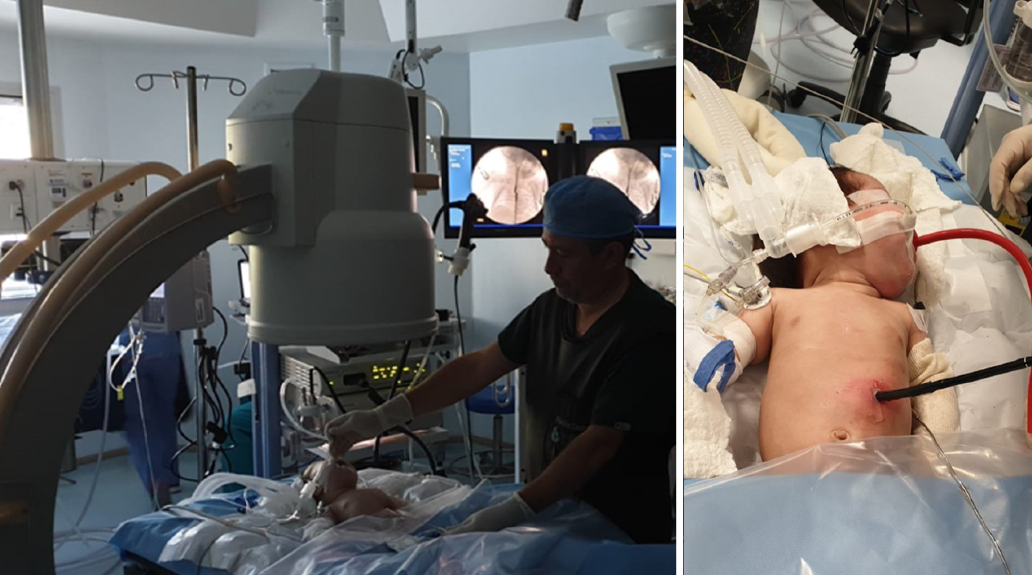
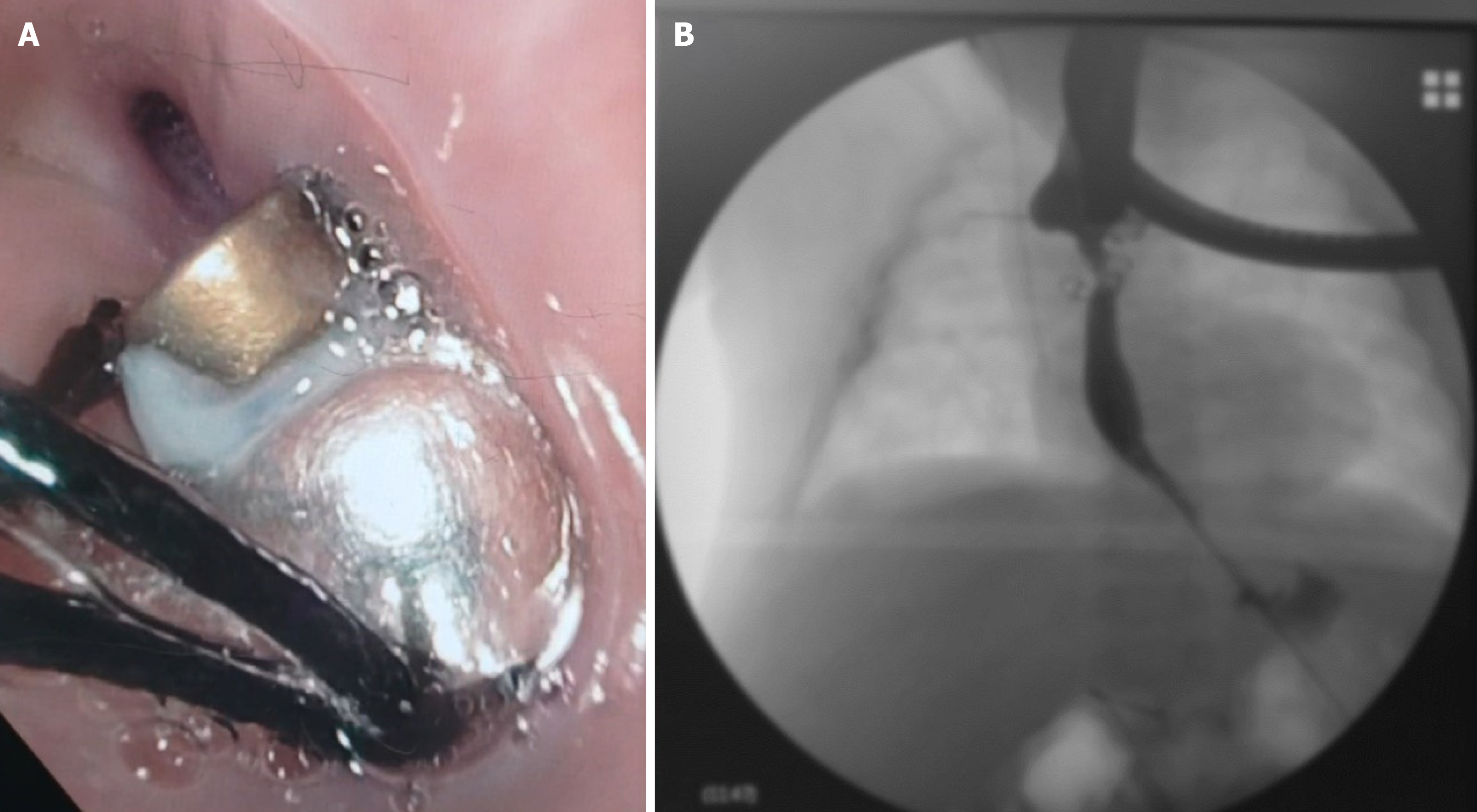
Figure 3 Placement of magnets in both esophageal pouches under endoscopic and fluoroscopic control.
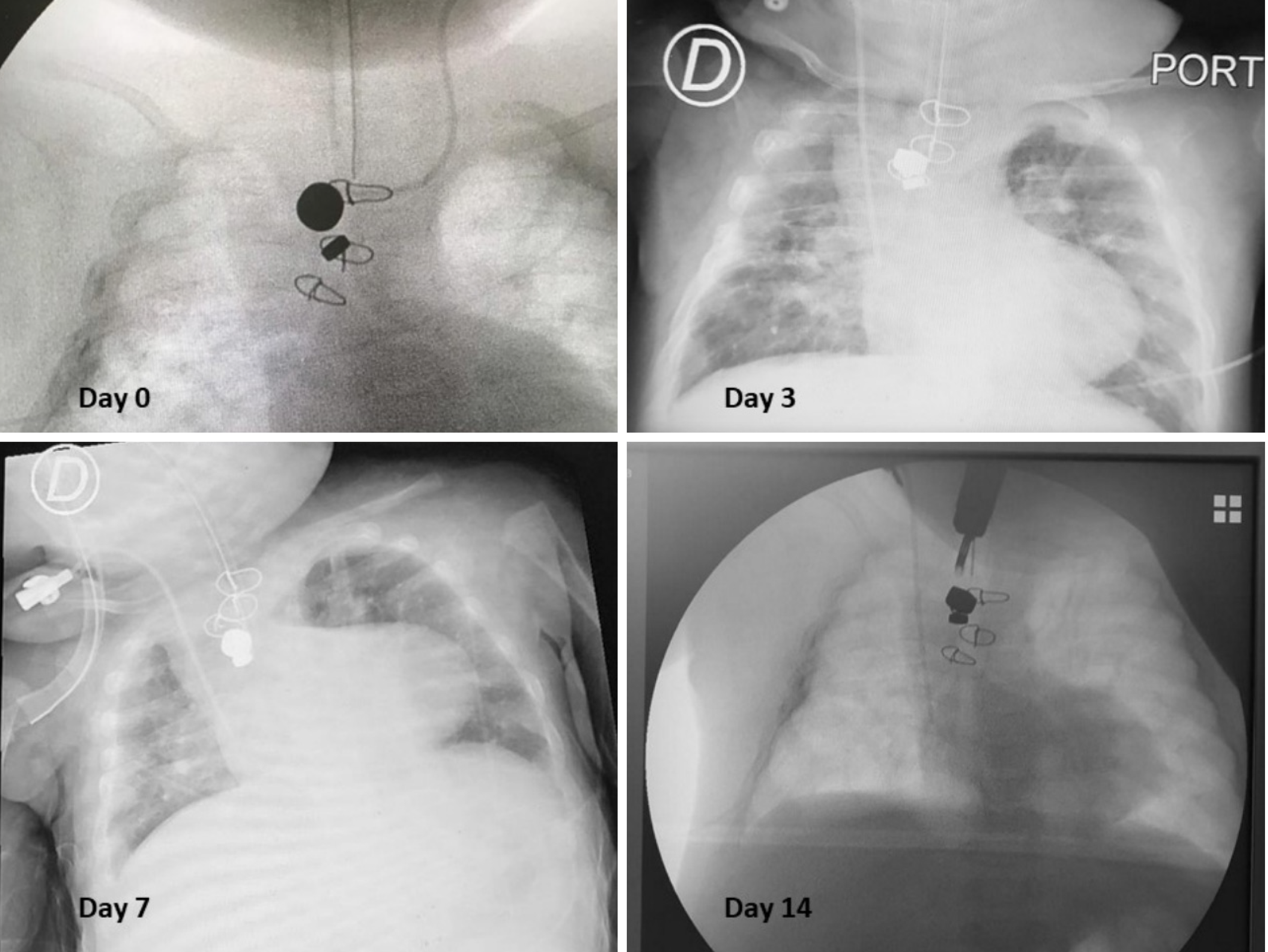
Figure 4 Radiological evolution while the magnets were in place.
Progressive mobilization closer together of the magnets is observed until they were completely joined.
Figure 5 Inmediate results after magnets removal.
A: Endoscopic removal of the magnets two weeks after placement; B: The esophagogram taken immediately after the procedure shows that the esophagus had been repermeabilized, but there is still some degree of stenosis at the anastomosis.
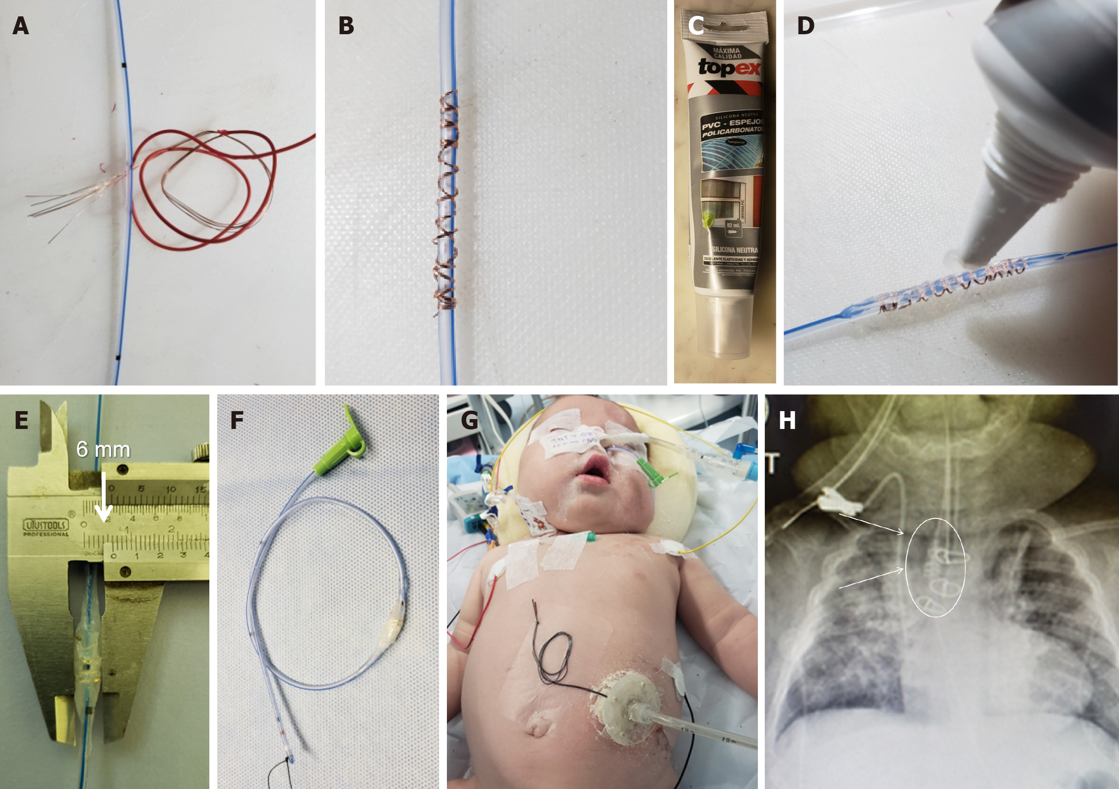
Figure 6 Custom-made dynamic stent.
A and B: A radio-opaque metal wire is coiled in a nasogastric tube according to stricture length, and is tailored to exceed the extent of the stricture by at least 2 cm to avoid displacement of the stent above or below the area of interest; C-E: The wire is covered with silicone until it reaches the desired thickness (6 mm in this case); F: A silk stitch is placed at the distal end of the probe for distal stent fixation; G, H: The dynamic stent is inserted in a way that is similar to a nasogastric tube, with the radio-opaque zone positioned in the stenotic area. It is secured with adhesive tape at the skin in two places (the probe near the nostril entrance and the silk stitch exiting through the gastrostomy).
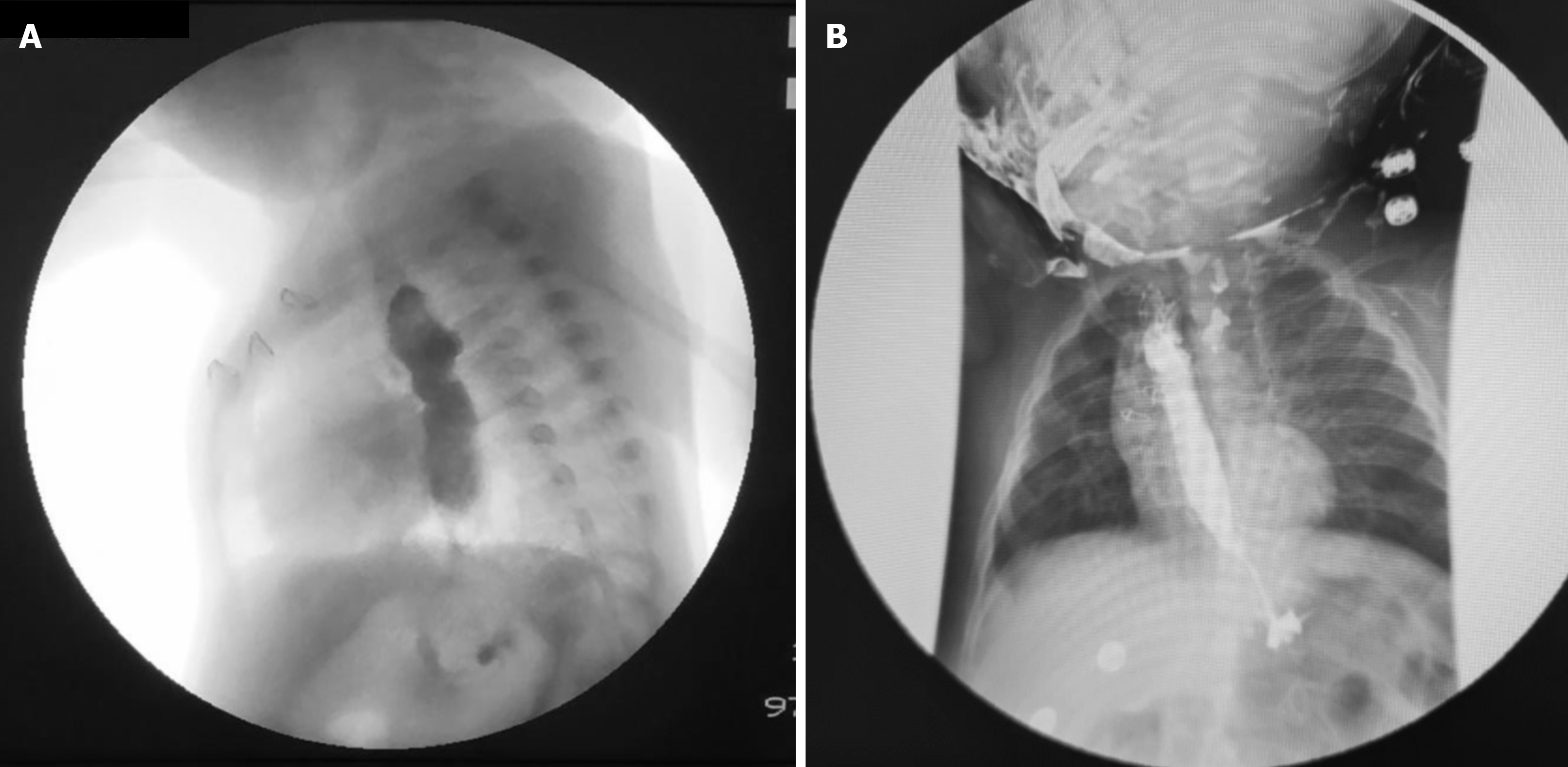
Figure 7 Absence of stricture recurrence after dynamic stent removal.
A: Esophagogram after inmediate removal of dynamic stent; B: Esophagogram 6 months later.
- Citation: Pérez-Bertólez S, Godoy-Lenz J. Primary repair of esophageal atresia Gross type C via thoracoscopic magnetic compression anastomosis: Is it the best option? World J Gastrointest Surg 2024; 16(5): 1474-1481
- URL: https://www.wjgnet.com/1948-9366/full/v16/i5/1474.htm
- DOI: https://dx.doi.org/10.4240/wjgs.v16.i5.1474


