Copyright
©The Author(s) 2024.
World J Gastrointest Surg. May 27, 2024; 16(5): 1395-1406
Published online May 27, 2024. doi: 10.4240/wjgs.v16.i5.1395
Published online May 27, 2024. doi: 10.4240/wjgs.v16.i5.1395
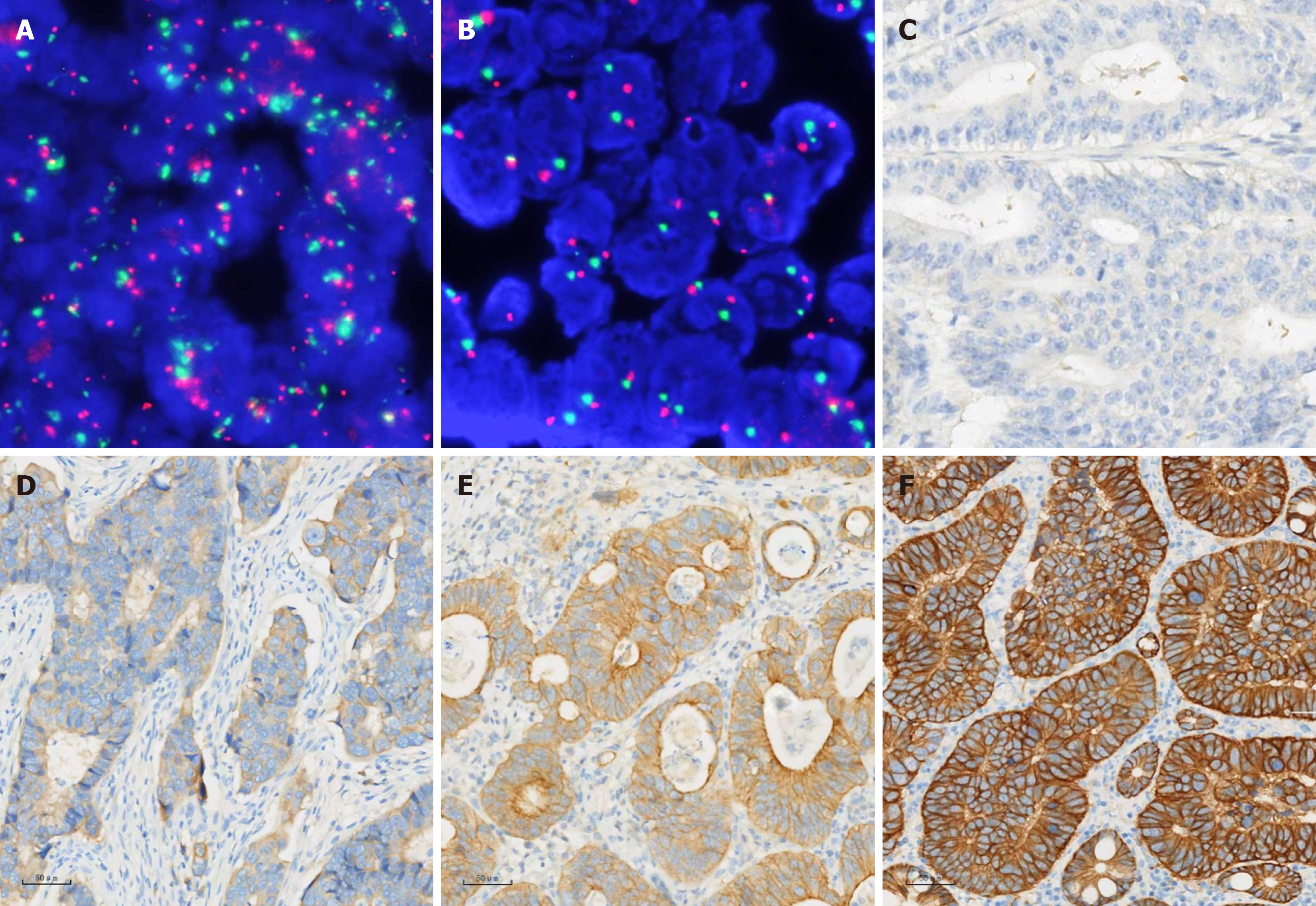
Figure 1 C-mesenchymal-epithelial transition factor immunohistochemistry (20 ×) and mesenchymal-epithelial transition factor fluorescence in situ hybridization (100 ×) results.
A: Mesenchymal-epithelial transition factor (MET) fluorescence in situ hybridization (FISH) positive (MET gene copy number ≥ 5 and MET/CEP7 < 2); B: MET FISH negative (MET gene copy number < 5 and MET/CEP7 < 2); C: Immunohistochemistry (IHC) 0; D: IHC 1+; E: IHC 2+; F: IHC 3+.
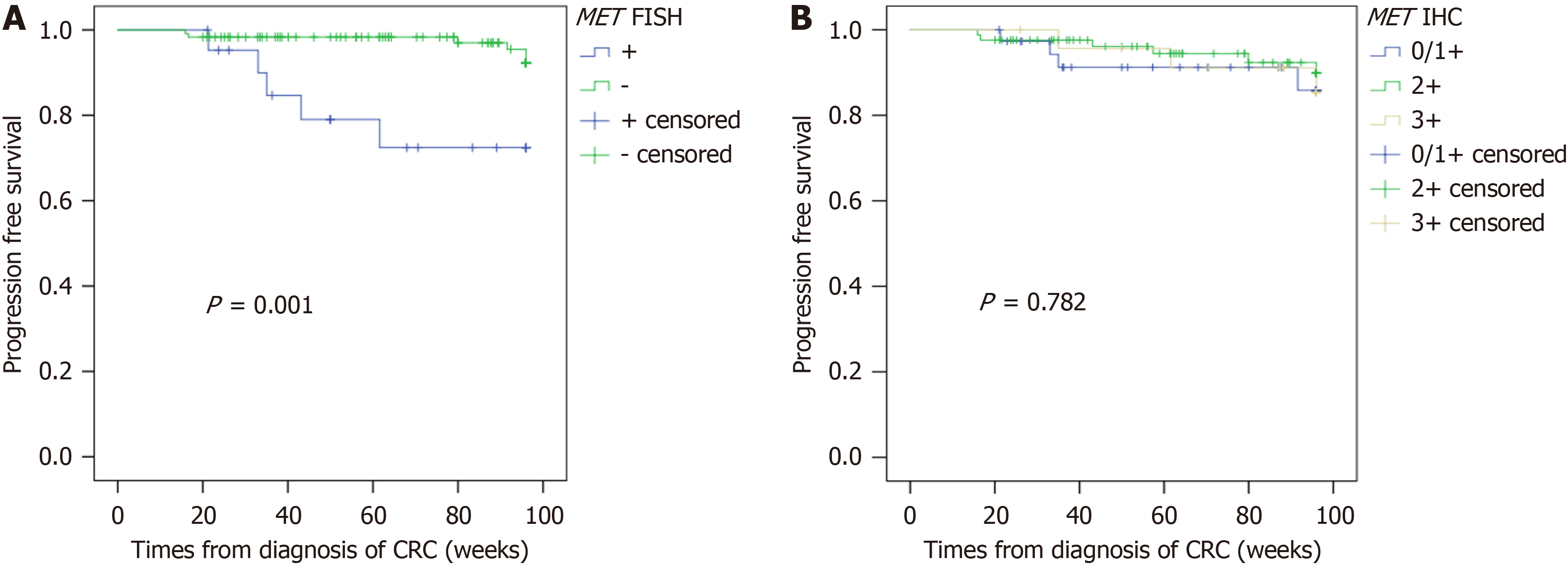
Figure 2 Kaplan-Meier curve of progression free survival for colorectal cancer patients in a two-year follow-up.
A: Progression free survival (PFS) in colorectal cancer (CRC) patient with amplified-mesenchymal-epithelial transition factor (MET) and non-amplified MET; B: PFS in CRC patient with different c-MET protein levels. MET: Mesenchymal-epithelial transition factor; FISH: Fluorescence in situ hybridization; IHC: Immunohistochemistry.
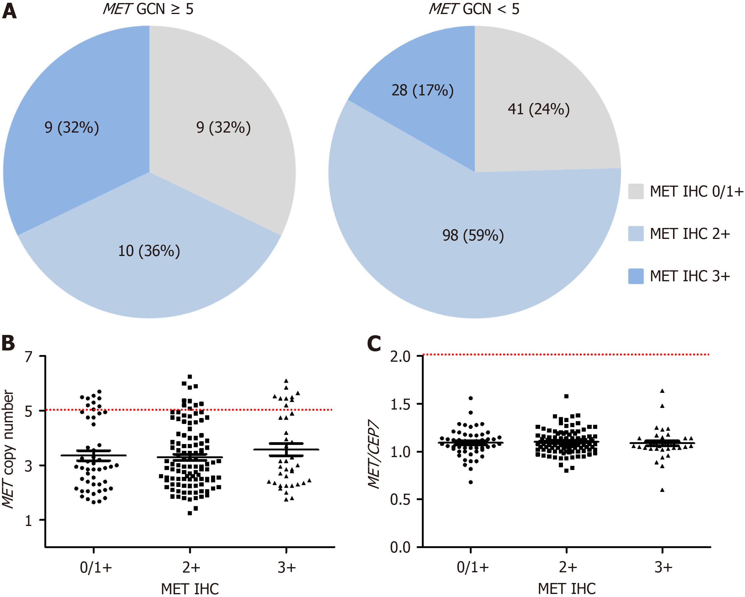
Figure 3 Comparison between mesenchymal-epithelial transition factor fluorescence in situ hybridization and immunohistochemistry results.
A: The c-mesenchymal-epithelial transition factor (MET) immunohistochemistry (IHC) status in MET fluorescence in situ hybridization positive and negative cases; B: The scatter plot representations of MET gene copy number in different c-MET IHC groups; C: The scatter plot representations of MET/CEP7 ratios in different c-MET IHC groups. MET: Mesenchymal-epithelial transition factor; GCN: Gene copy number; IHC: Immunohistochemistry.
- Citation: Yu QX, Fu PY, Zhang C, Li L, Huang WT. Mesenchymal-epithelial transition factor amplification correlates with adverse pathological features and poor clinical outcome in colorectal cancer. World J Gastrointest Surg 2024; 16(5): 1395-1406
- URL: https://www.wjgnet.com/1948-9366/full/v16/i5/1395.htm
- DOI: https://dx.doi.org/10.4240/wjgs.v16.i5.1395


