Copyright
©The Author(s) 2024.
World J Gastrointest Surg. May 27, 2024; 16(5): 1336-1343
Published online May 27, 2024. doi: 10.4240/wjgs.v16.i5.1336
Published online May 27, 2024. doi: 10.4240/wjgs.v16.i5.1336
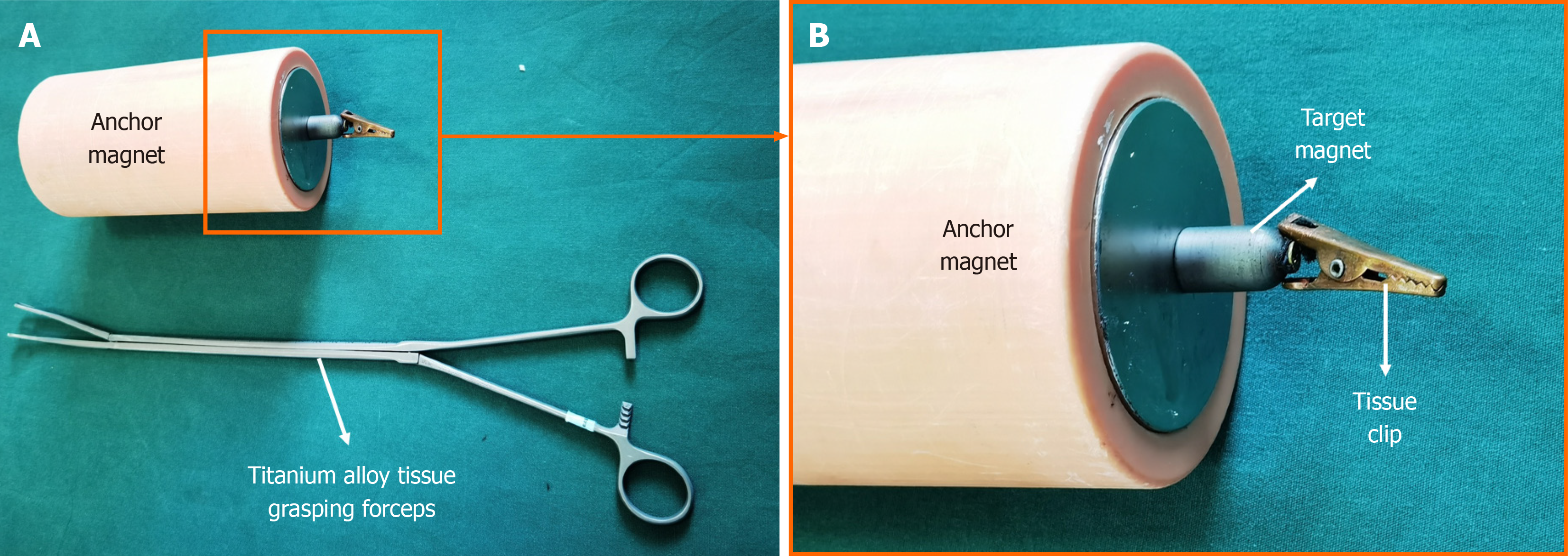
Figure 1 Y-Z magnetic anchor devices.
A: Anchor magnet and the titanium alloy tissue grasping forceps; B: Magnetic grasping apparatus.
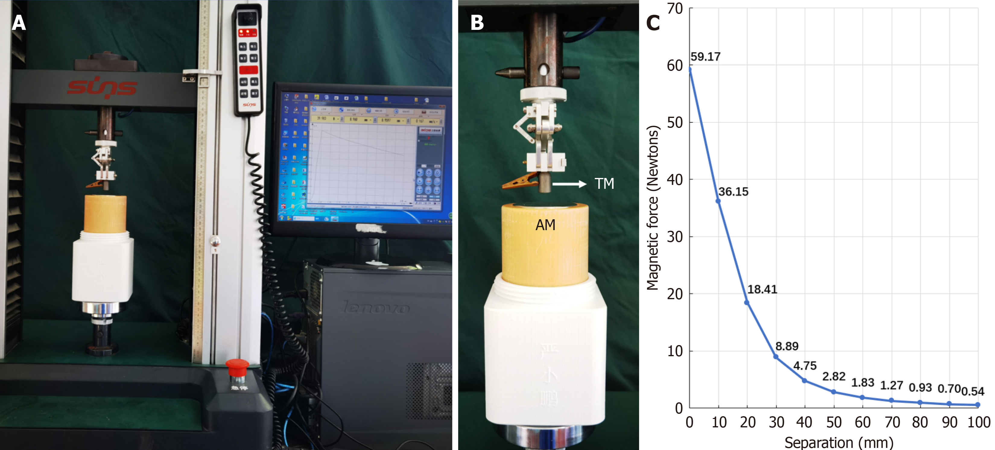
Figure 2 Magnetism test of the Y-Z magnetic anchor devices.
A: Magnetism test equipment; B: The anchor magnet and target magnet in the test state; C: The magnetic force-displacement curve of the anchor and target magnet. AM: Anchor magnet; TM: Target magnet.
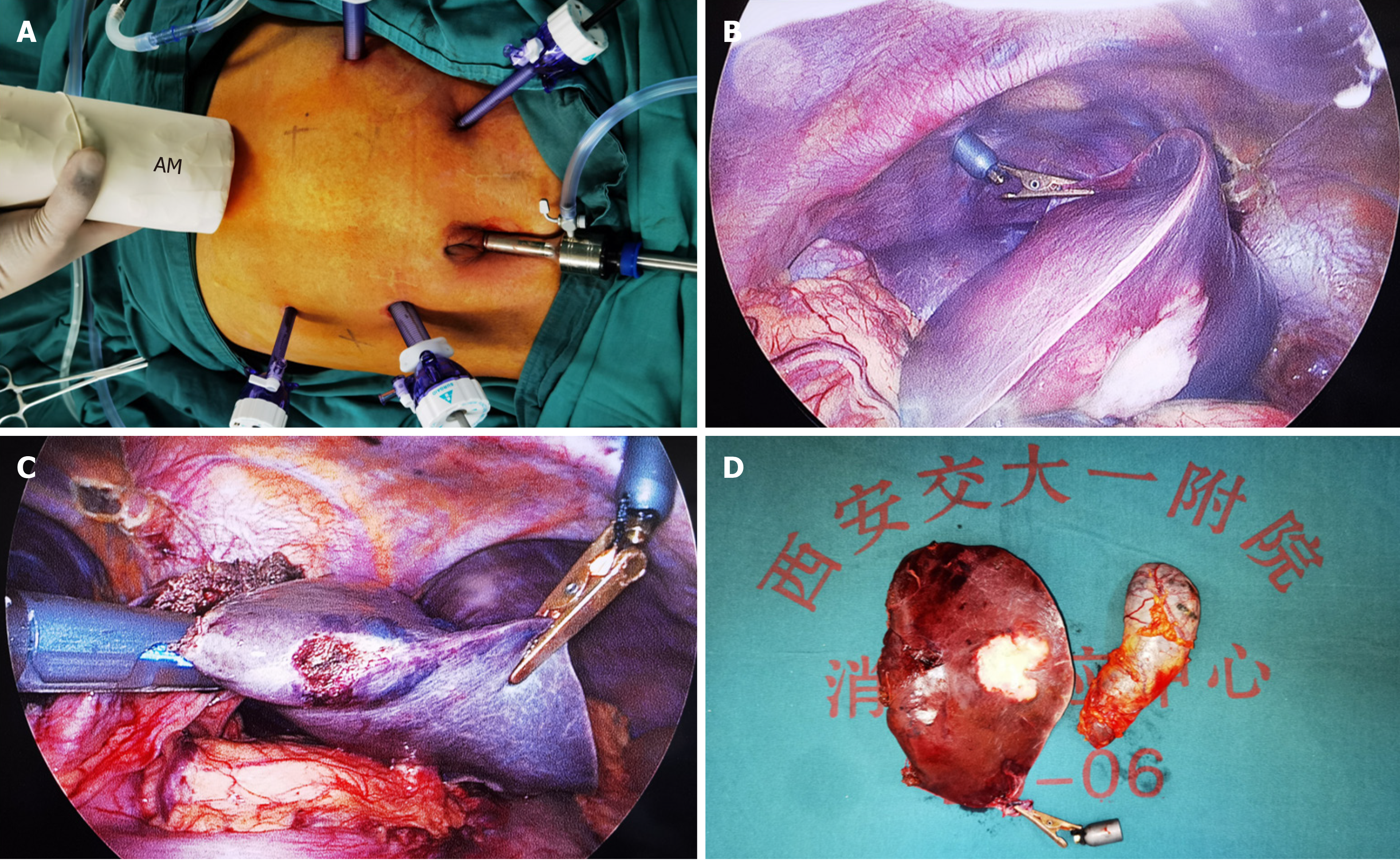
Figure 3 Magnetic anchor-assisted 5-port left lateral hepatic lobectomy operation procedure (patient 2).
A: The layout of the abdominal wall port and the position of the anchor magnet; B: The magnetic anchoring device pulling the left lateral lobe of the liver to reveal the tumor lesion and the hepatogastric ligament; C: The left lateral lobe of the liver is shown under the magnetic anchoring device, which is transected at the hepatic pedicle of the left lateral lobe of the liver with a linear cutting and closure device; D: The left lateral hepatic lobe and gallbladder specimens that are resected. AM: Anchor magnet.
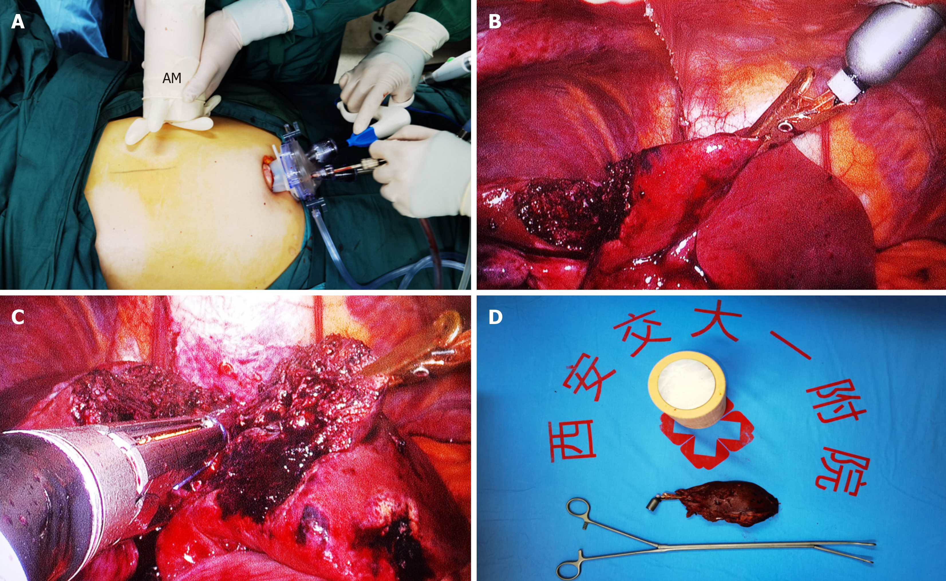
Figure 4 Magnetic anchoring-assisted transumbilical single-port laparoscopic left lateral hepatic lobectomy process (patient 5).
A: The subumbilical single-port and the anchor magnet; B: The Y-Z magnetic anchor devices pulling the left lateral lobe of the liver to reveal the liver section; C: The linear cutting and closure device was used to cut off segment II and part of segment III of the left lateral lobe of the liver; D: The resected segment II and part of segment III of the left lateral lobe of the liver. AM: Anchor magnet.
- Citation: Zhang MM, Bai JG, Zhang D, Tao J, Geng ZM, Li ZQ, Ren YX, Zhang YH, Lyu Y, Yan XP. Clinical feasibility of laparoscopic left lateral segment liver resection with magnetic anchor technique: The first clinical study from China. World J Gastrointest Surg 2024; 16(5): 1336-1343
- URL: https://www.wjgnet.com/1948-9366/full/v16/i5/1336.htm
- DOI: https://dx.doi.org/10.4240/wjgs.v16.i5.1336


