Copyright
©The Author(s) 2024.
World J Gastrointest Surg. May 27, 2024; 16(5): 1311-1319
Published online May 27, 2024. doi: 10.4240/wjgs.v16.i5.1311
Published online May 27, 2024. doi: 10.4240/wjgs.v16.i5.1311
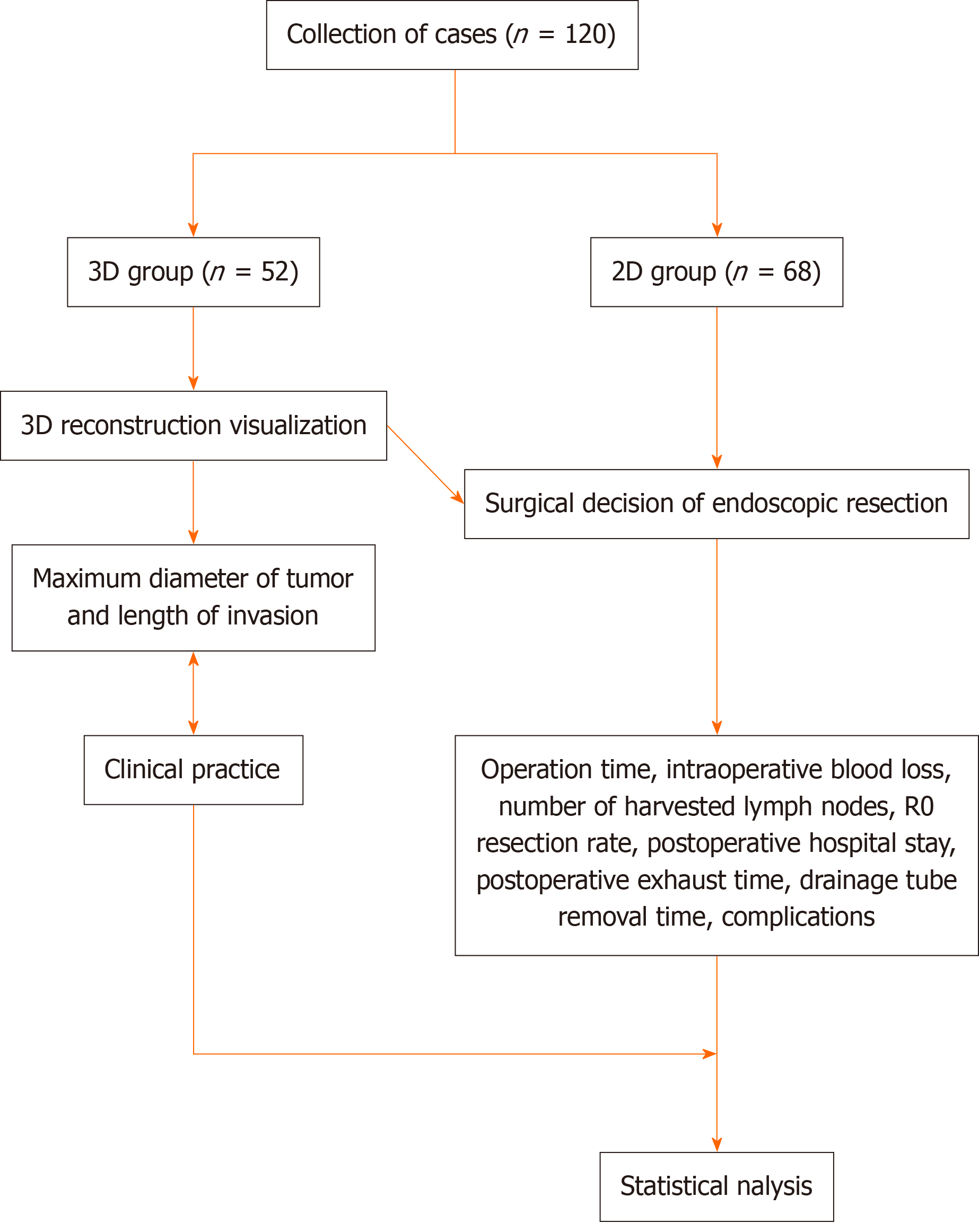
Figure 1 Flowchart of the research scheme.
3D: Three-dimensional; 2D: Two-dimensional.
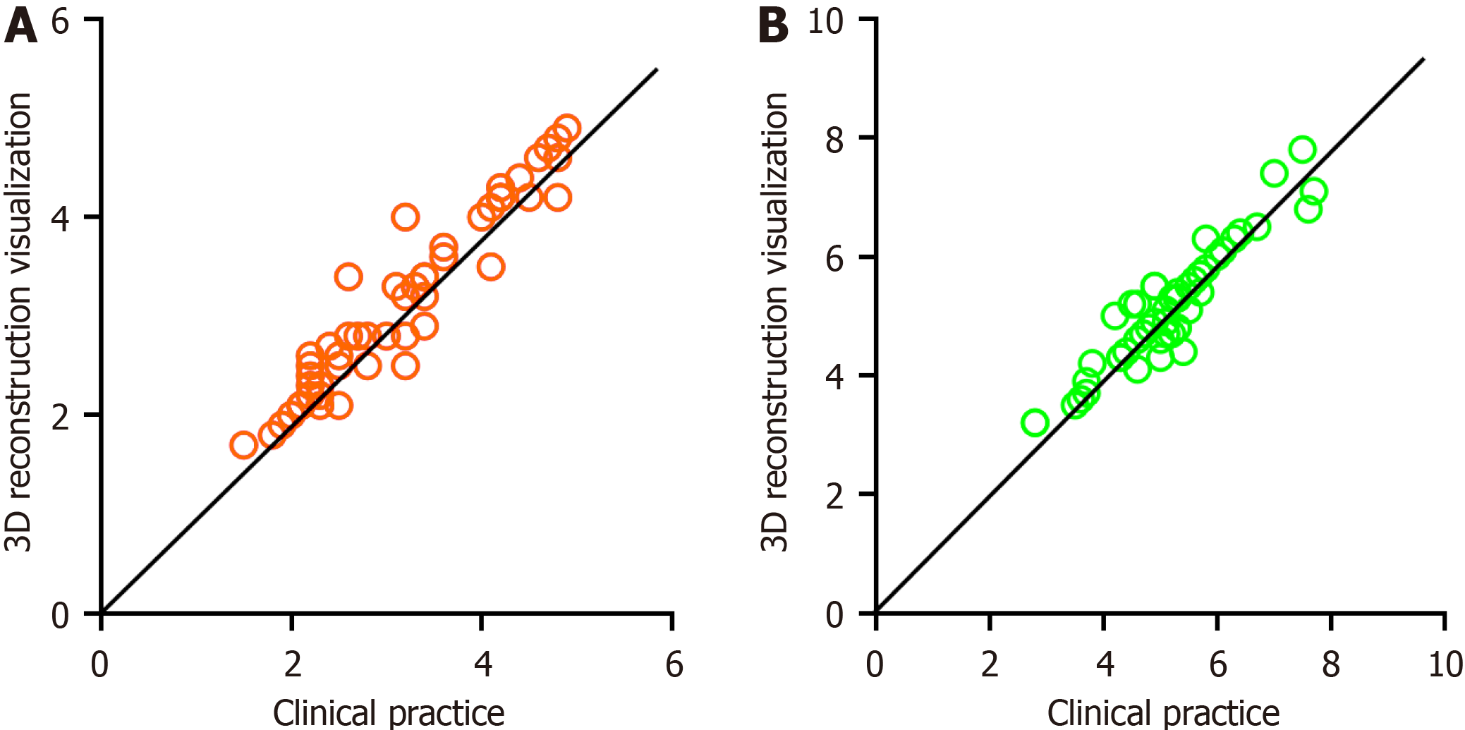
Figure 2 Actual tumor conditions under three-dimensional reconstruction visualization and in clinical practice.
A: Maximum tumor diameter; B: infiltration length. 3D: Three-dimensional; 2D: Two-dimensional.
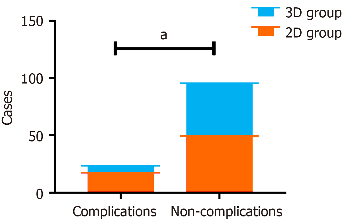
Figure 3 Stacking diagram of the proportion of complications in the three-dimensional and two-dimensional groups.
aP < 0.05. 3D: Three-dimensional; 2D: Two-dimensional.
- Citation: Guo D, Zhu XY, Han S, Liu YS, Cui DP. Evaluating the use of three-dimensional reconstruction visualization technology for precise laparoscopic resection in gastroesophageal junction cancer. World J Gastrointest Surg 2024; 16(5): 1311-1319
- URL: https://www.wjgnet.com/1948-9366/full/v16/i5/1311.htm
- DOI: https://dx.doi.org/10.4240/wjgs.v16.i5.1311


