Copyright
©The Author(s) 2024.
World J Gastrointest Surg. Apr 27, 2024; 16(4): 1109-1120
Published online Apr 27, 2024. doi: 10.4240/wjgs.v16.i4.1109
Published online Apr 27, 2024. doi: 10.4240/wjgs.v16.i4.1109
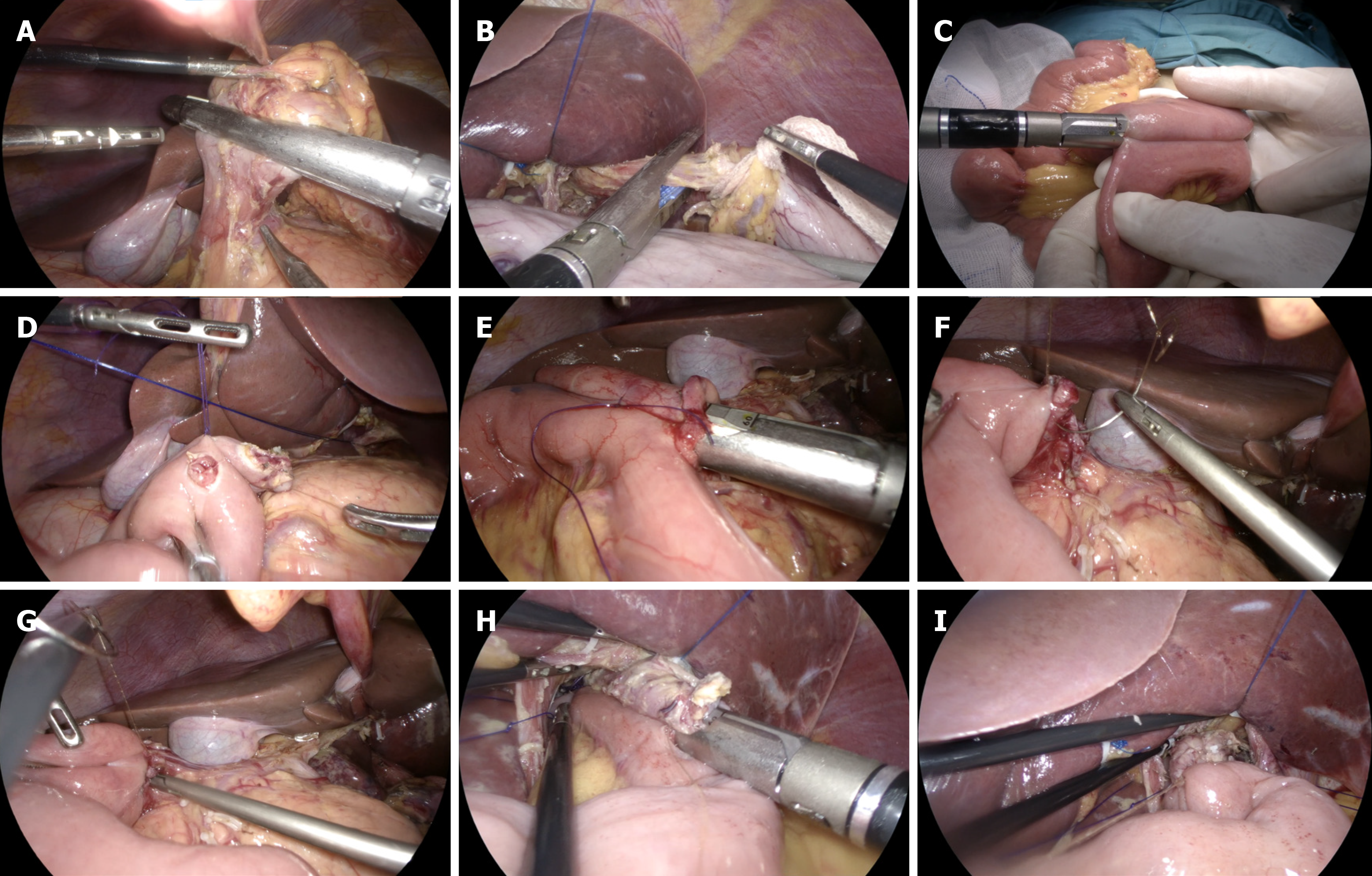
Figure 1 Total laparoscopic radical total gastrectomy + jejunal interposition double-tract reconstruction.
A: After cleaning the upper and lower pyloric lymph nodes, the duodenum was severed using a linear cutting stapler; B: Disconnection of esophagus; C: The Jejunal side-to-side anastomosis was completed by a linear cutting stapler; D: Duodenum and jejunum openings; E: Linear cutting stapler to complete duodenum-jejunum side-to-side anastomosis; F: The common opening of duodenum jejunum anastomosis was sutured by barbed suture; G: Suture the duodenal stump with barbed suture; H: Linear cutting stapler to complete esophageal jejunum overlap anastomosis; I: The barbed suture was continuously sutured to close the common opening of esophagus jejunum.
Figure 2 Two groups of patients with postoperative white blood cells, neutrophils and platelets change line chart.
A: White blood cells change line chart; B: Neutrophils change line chart; C: Platelets change line chart. DTR: Double-tract reconstruction; RY: Roux-en-Y reconstruction; WBC: White blood cells; NEU: Neutrophils; PLT: Platelets; Pre: Preoperative; Pos 1d: The first day after surgery; Pos 3d: The third day after surgery; Pos 5d: The fifth day after surgery.
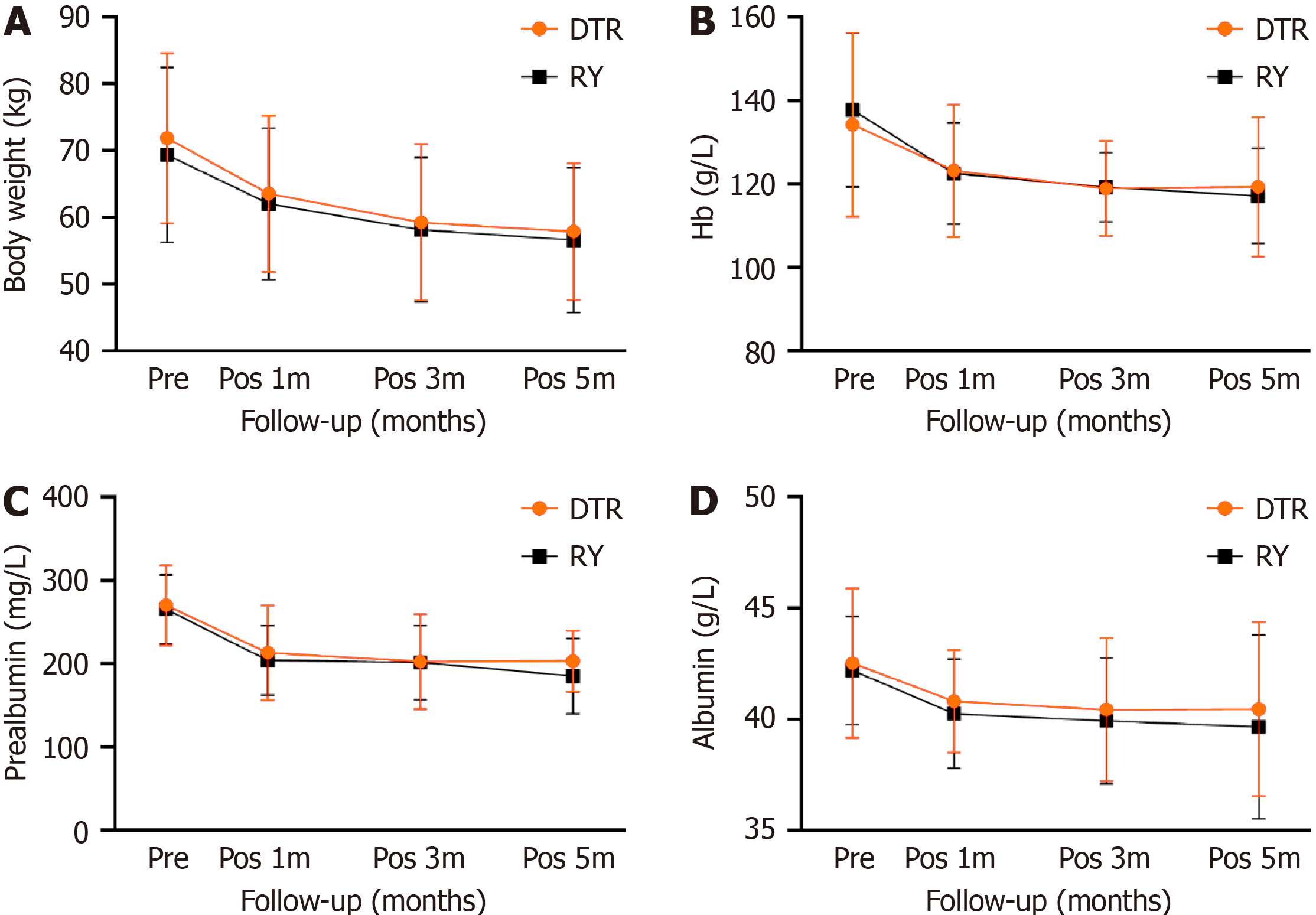
Figure 3 Line chart showing the body weight, prealbumin, albumin, and hemoglobin changes of the two groups of patients.
A: Body weight change line chart; B: Hemoglobin change line chart; C: Prealbumin change line chart; D: Albumin change line chart. DTR: Double-tract reconstruction; RY: Roux-en-Y reconstruction; Hb: Hemoglobin; Pre: Preoperative; Pos 1m: At 1 month after operation; Pos 3m: At 3 months after operation; Pos 6m: At 6 months after operation.
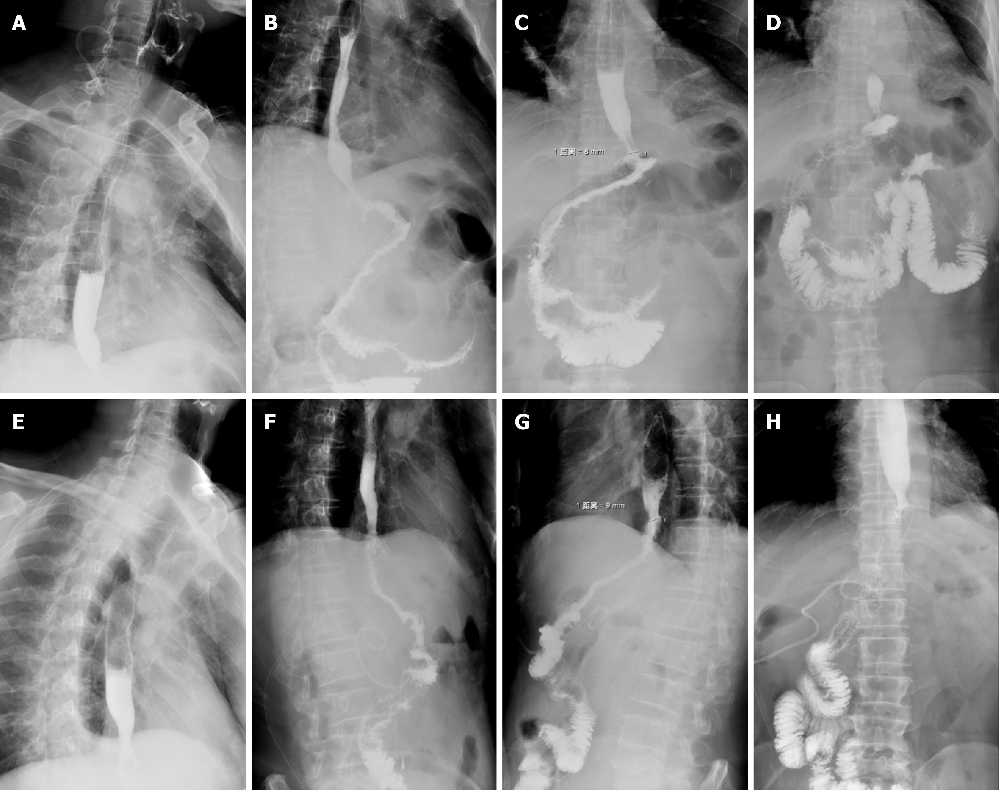
Figure 4 Postoperative angiography of the two groups of patients.
A-D: Double-tract reconstruction: Imaging of contrast agent in esophagus (A), the contrast agent passed through the esophagojejunal anastomosis smoothly (B), the contrast agent successfully passed through the duodenal jejunum anastomosis (C), and imaging of contrast agent in distal jejunum (D); E-H: Roux-en-Y reconstruction: Imaging of contrast agent in esophagus (E), the contrast agent passed through the esophagojejunal anastomosis smoothly (F), imaging of contrast agent in proximal jejunum (G), and imaging of contrast agent in distal jejunum (H).
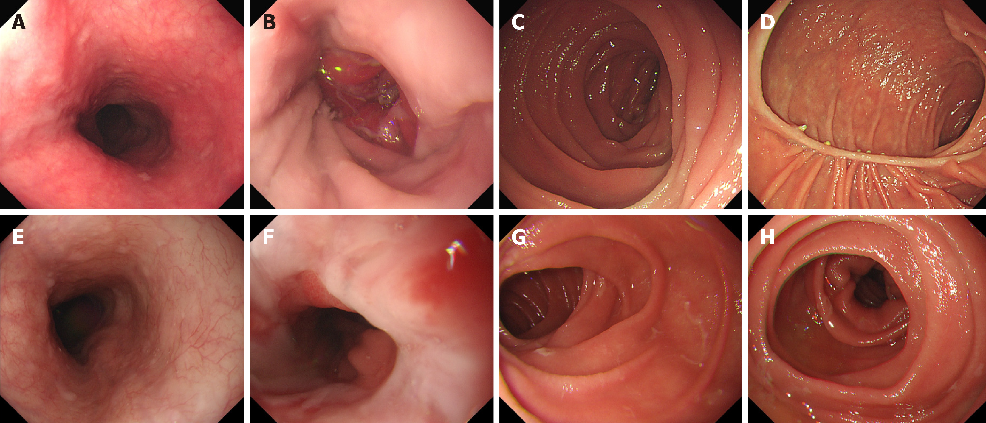
Figure 5 Postoperative gastroscopy results of the two groups of patients.
A-D: Double-tract reconstruction: Image of esophagus under gastroscope (A), image of esophagojejunal anastomosis under gastroscope (B), image of proximal jejunum under gastroscope (C), and image of duodenal jejunum anastomosis under gastroscope (D); E-H: Roux-en-Y reconstruction: Image of esophagus under gastroscope (E), image of esophagojejunal anastomosis under gastroscope (F), image of proximal jejunum under gastroscope (G), and image of distal jejunum under gastroscope (H).
- Citation: Dong TX, Wang D, Zhao Q, Zhang ZD, Zhao XF, Tan BB, Liu Y, Liu QW, Yang PG, Ding PA, Zheng T, Li Y, Liu ZJ. Comparative analysis of two digestive tract reconstruction methods in total laparoscopic radical total gastrectomy. World J Gastrointest Surg 2024; 16(4): 1109-1120
- URL: https://www.wjgnet.com/1948-9366/full/v16/i4/1109.htm
- DOI: https://dx.doi.org/10.4240/wjgs.v16.i4.1109


