Copyright
©The Author(s) 2024.
World J Gastrointest Surg. Mar 27, 2024; 16(3): 955-965
Published online Mar 27, 2024. doi: 10.4240/wjgs.v16.i3.955
Published online Mar 27, 2024. doi: 10.4240/wjgs.v16.i3.955
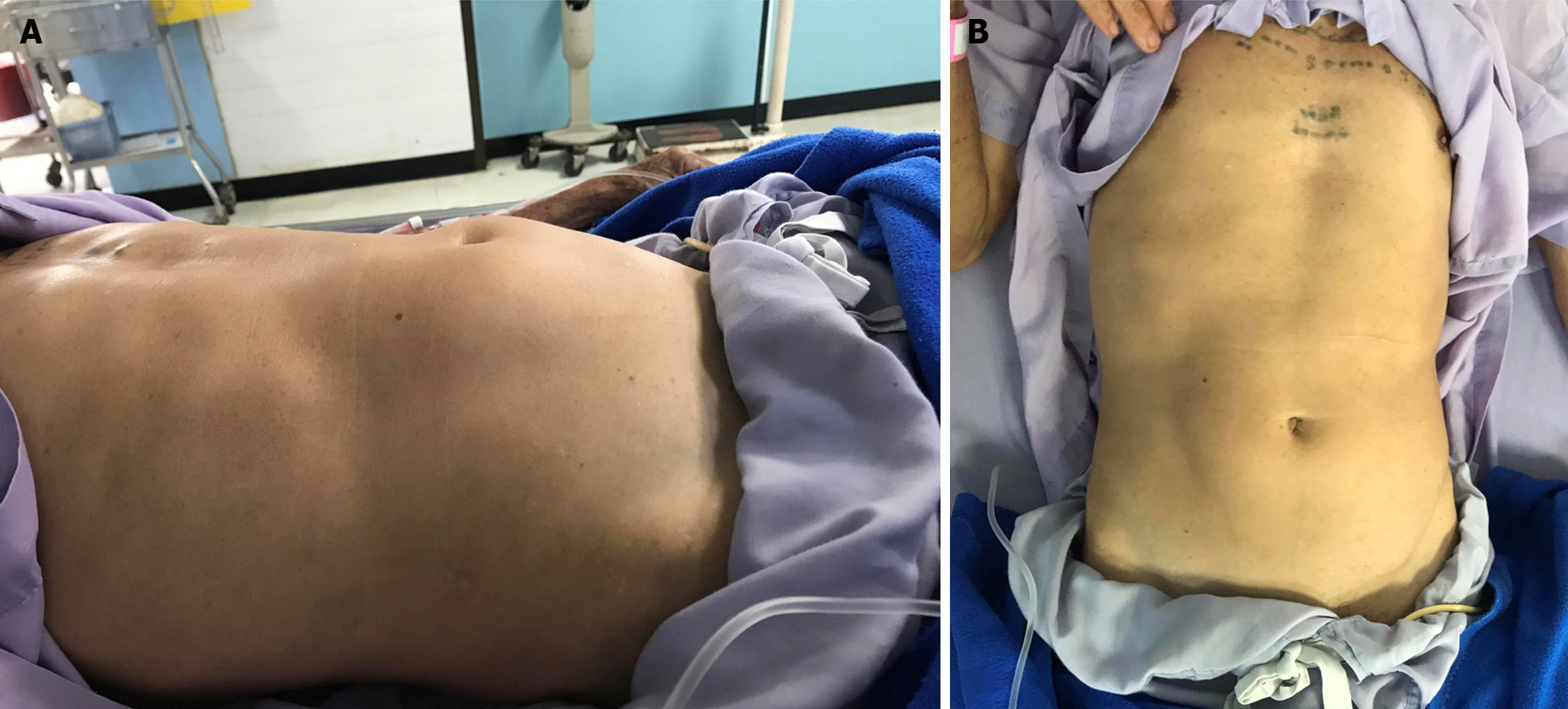
Figure 1 Physical exam of bulging mid-abdomen.
A: Lateral view of abdomen; B: Bird eye view of abdomen.
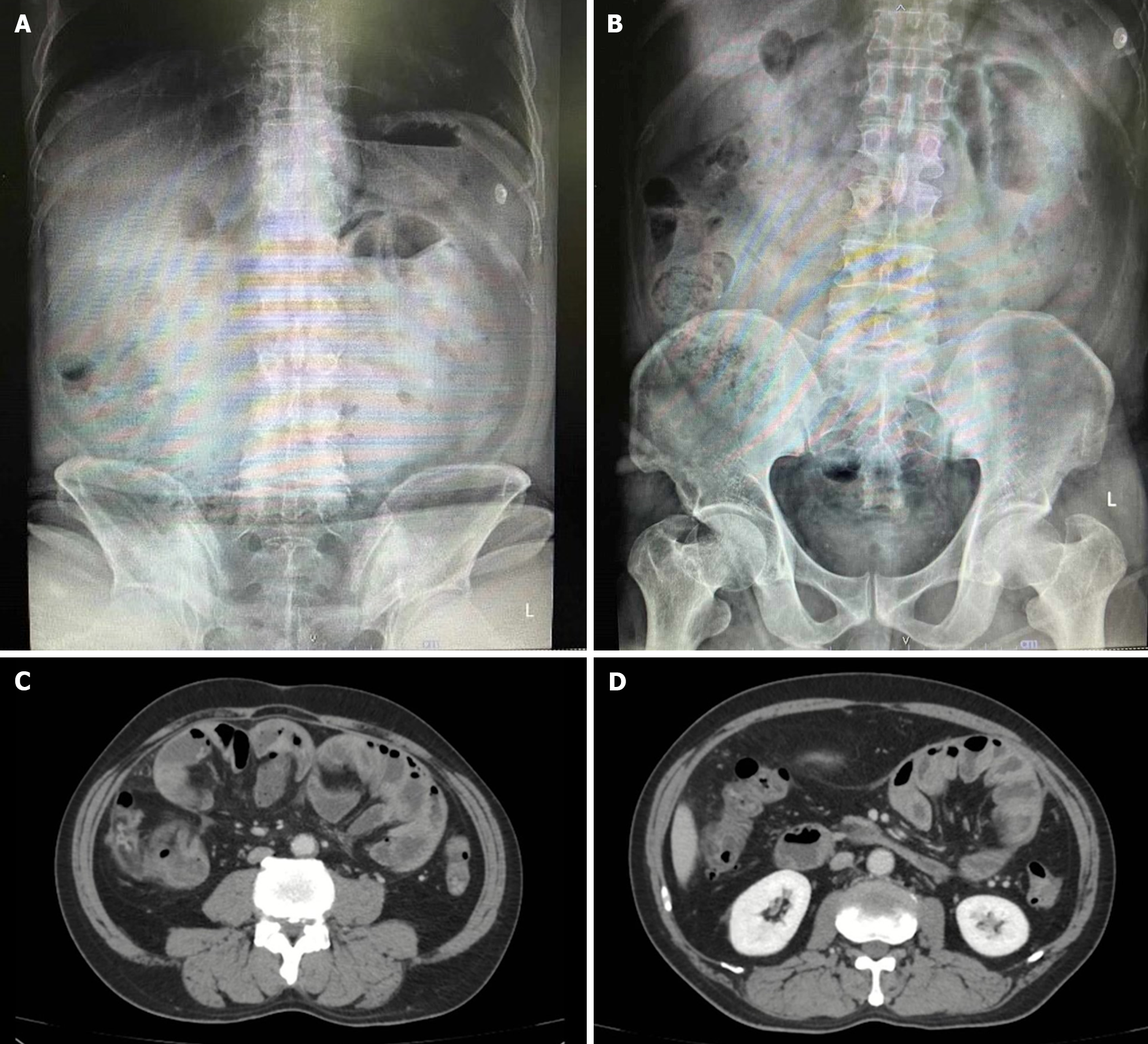
Figure 2 Abdominal X-ray and computed tomography scan whole abdomen with contrast.
A: Upright view; B: Supine view; C and D: Computed tomography scan of enhancing peritoneum (cocoon-like) without small bowel dilatation.
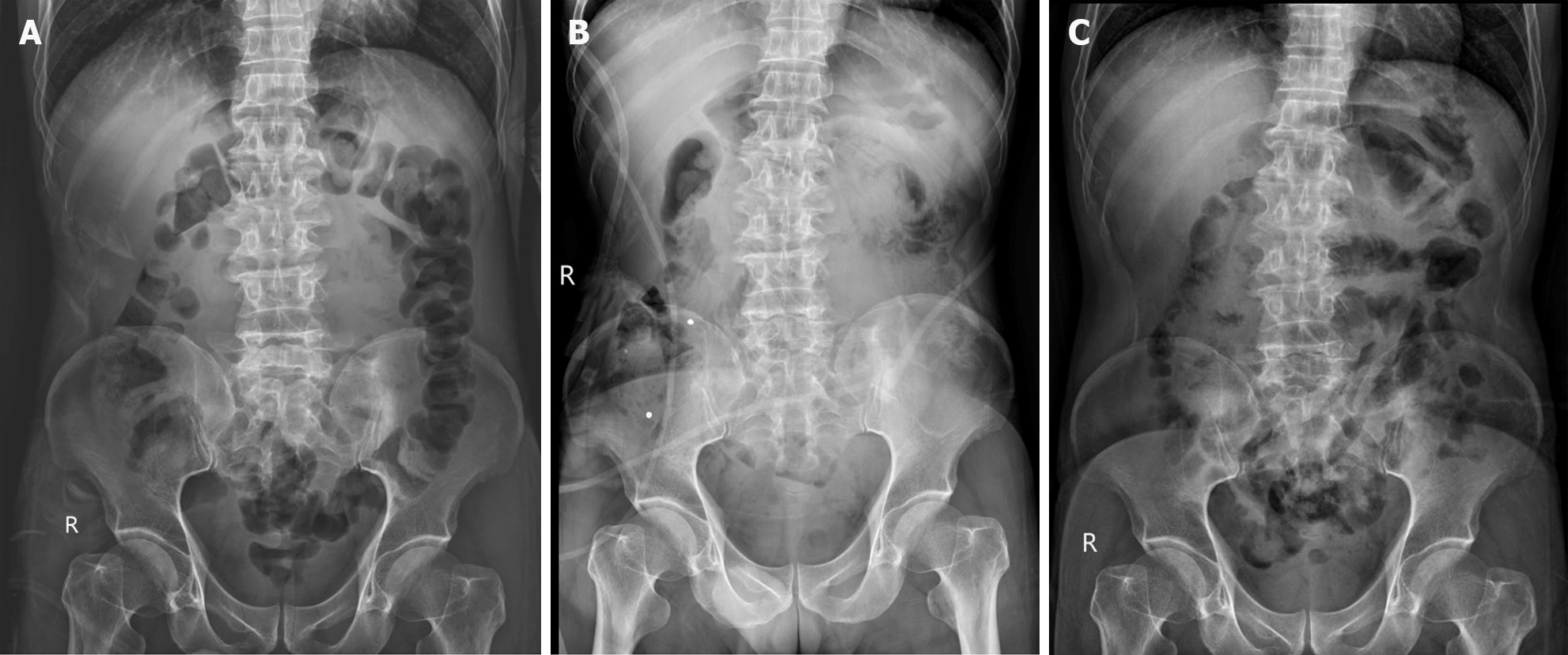
Figure 3 Abdominal X-rays.
A: Gasless small bowel from X-ray in 1st visit; B: Small bowel devoid of gas observed on the fifth day after the completion of a colonoscopy procedure; C: Disappearance of gasless small bowel after operation.
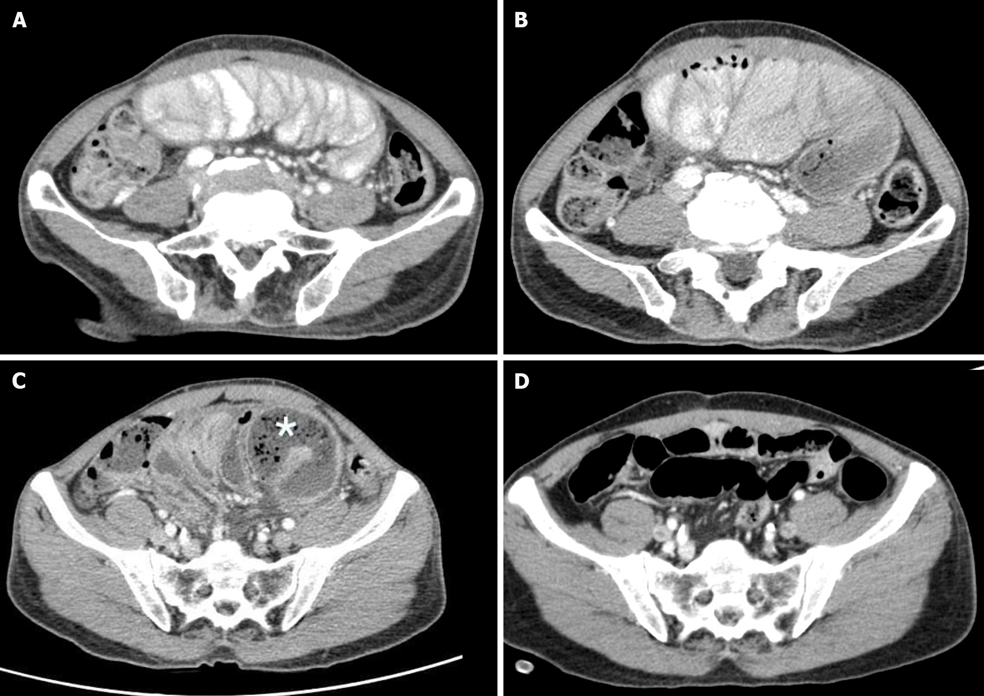
Figure 4 Computed tomography scan whole abdomen with contrast.
A and B: Stacking small bowel loops inside a thick membrane-like sac; C: Small bowel feces sign (asterisk); D: Normal alignment of the small bowel after operation.
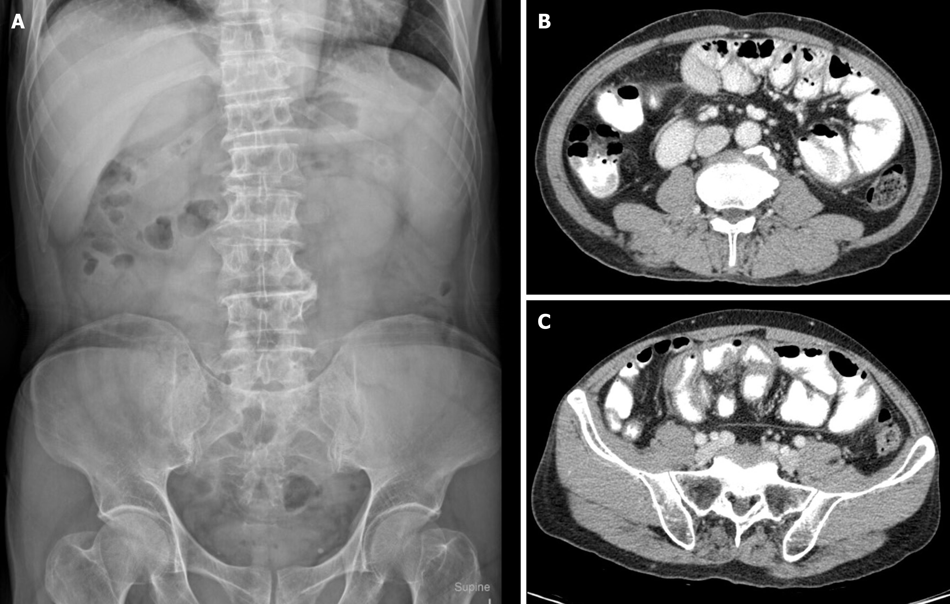
Figure 5 Abdominal X-ray and computed tomography whole abdomen with oral and IV contrast.
A: Gasless small bowel; B and C: Stacking small bowel loops inside a thick membrane-like sac.
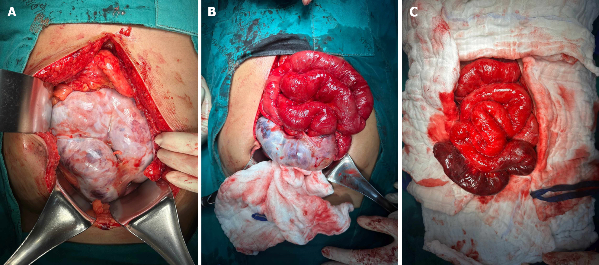
Figure 6 Intraoperative finding of encapsulated of intestine.
A: Entire intestine with dense fibrous tissue; B: Partial resection of dense fibrous tissue; C: After complete resection of all fibrous tissue with small bowel necrosis.
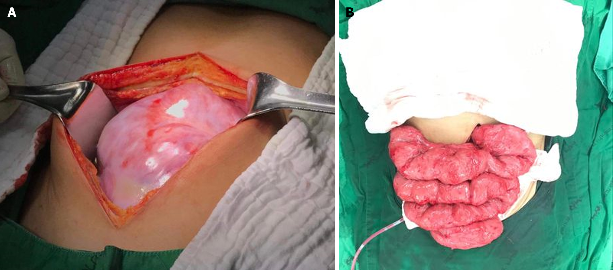
Figure 7 Intraoperative images.
A: Fibrous sheath encapsulated small bowel; B: Small bowel after complete adhesiolysis.
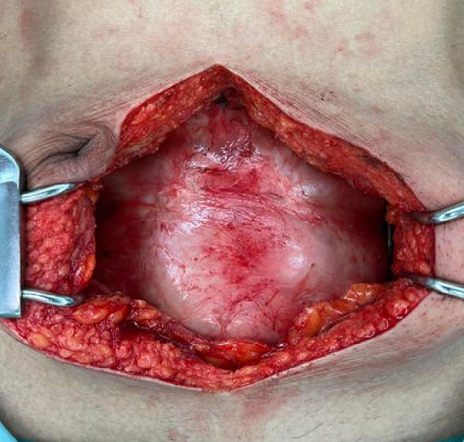
Figure 8
Intraoperative finding.
- Citation: Vipudhamorn W, Juthasilaparut T, Sutharat P, Sanmee S, Supatrakul E. Abdominal cocoon syndrome-a rare culprit behind small bowel ischemia and obstruction: Three case reports. World J Gastrointest Surg 2024; 16(3): 955-965
- URL: https://www.wjgnet.com/1948-9366/full/v16/i3/955.htm
- DOI: https://dx.doi.org/10.4240/wjgs.v16.i3.955


