Copyright
©The Author(s) 2024.
World J Gastrointest Surg. Feb 27, 2024; 16(2): 628-634
Published online Feb 27, 2024. doi: 10.4240/wjgs.v16.i2.628
Published online Feb 27, 2024. doi: 10.4240/wjgs.v16.i2.628
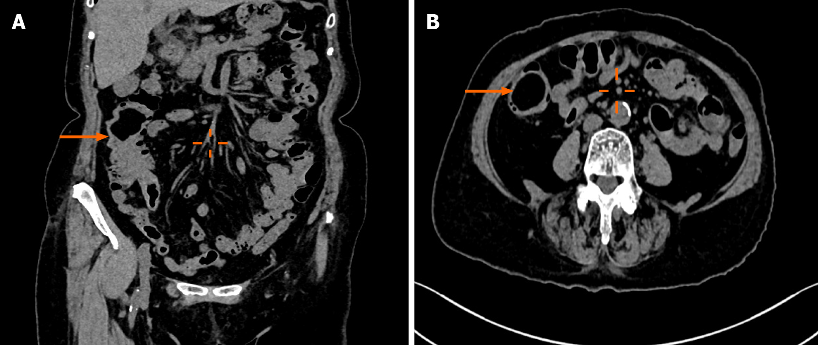
Figure 1 Contrast-enhanced computed tomography of the abdomen.
A: Partial invagination of the ascending colon suggests intussusception (orange arrow); B: The mass at the terminal ileum (orange arrow).
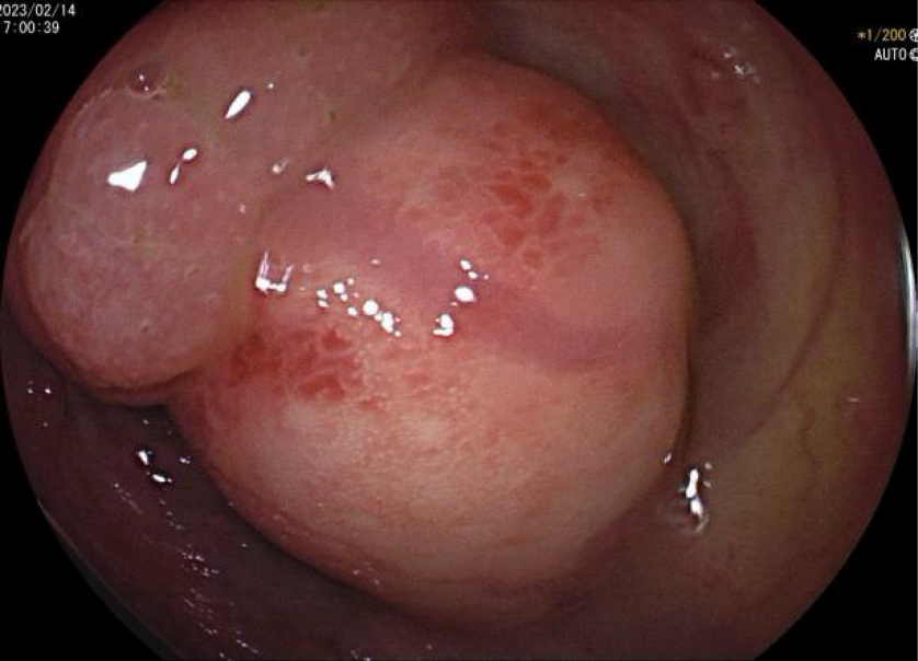
Figure 2 Colonoscopy examination.
Submucosal elevation 6 cm from the ileocecal valve consists of two distinct parts with a clearly visible demarcation line.
Figure 3 Modified endoscopic submucosal dissection procedure.
A: Endoscopic images showing the process of endoscopic submucosal dissection (ESD) for the tumor; B: Endoscopic images showing the purse-string suture closure of the excised tumor site after ESD; C: The resected mass.
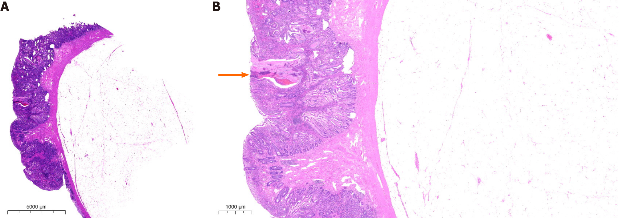
Figure 4 Histopathological examination.
A: Microscopic examination revealed a benign collision tumor (lipoma combined with juvenile polyp); B: The signs of angiodysplasia and bleeding on the surface of the lesion (orange arrow).
- Citation: Wu YQ, Wang HY, Shao MM, Xu L, Jiang XY, Guo SJ. Ileal collision tumor associated with gastrointestinal bleeding: A case report and review of literature. World J Gastrointest Surg 2024; 16(2): 628-634
- URL: https://www.wjgnet.com/1948-9366/full/v16/i2/628.htm
- DOI: https://dx.doi.org/10.4240/wjgs.v16.i2.628


