Copyright
©The Author(s) 2024.
World J Gastrointest Surg. Feb 27, 2024; 16(2): 622-627
Published online Feb 27, 2024. doi: 10.4240/wjgs.v16.i2.622
Published online Feb 27, 2024. doi: 10.4240/wjgs.v16.i2.622
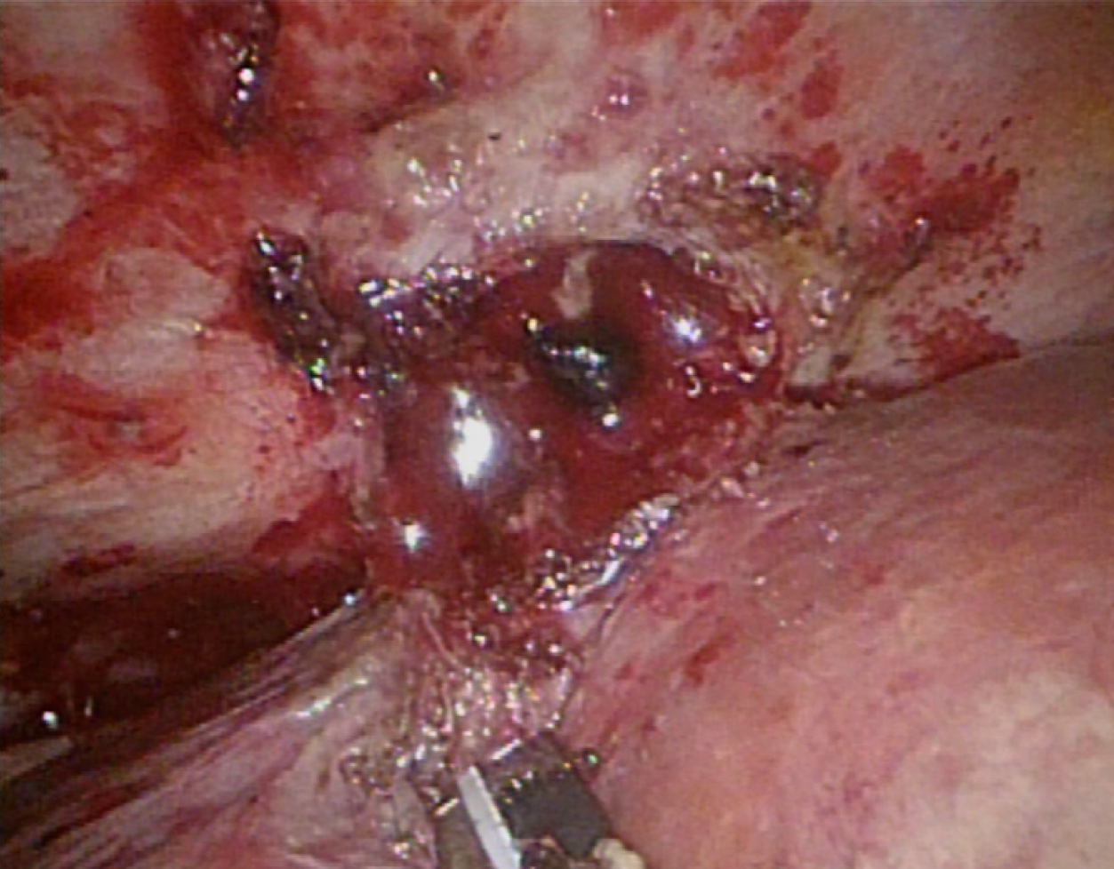
Figure 1 Cholelith-like mass with abscess on the surface of the S7 segment of the liver.
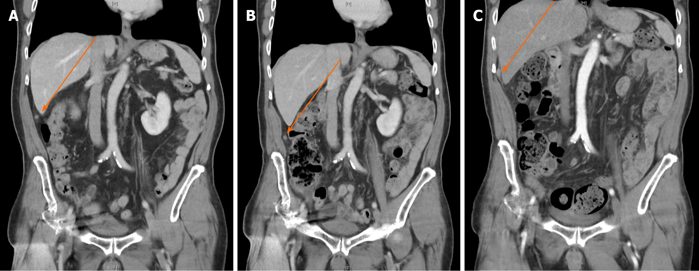
Figure 2 Suspected local recurrence or metastasis on abdominal computed tomography scan.
A: August 2022; B: December 2022; C: March 2023.
Figure 3 Suspected tumor seeding via whole-body positron emission tomography.
Figure 4 Histological examination of the partial resection resection omentum.
Brownish foreign body substances were present and surrounded by purulent inflammatory cells and foreign body giant cells. Brownish the brownish foreign material was stone.
- Citation: Huang CK, Lu RH, Chen CC, Chen PC, Hsu WC, Tsai MJ, Ting CT. Spilled gallstone mimicking intra-abdominal seeding of gallbladder adenocarcinoma: A case report. World J Gastrointest Surg 2024; 16(2): 622-627
- URL: https://www.wjgnet.com/1948-9366/full/v16/i2/622.htm
- DOI: https://dx.doi.org/10.4240/wjgs.v16.i2.622


