Copyright
©The Author(s) 2024.
World J Gastrointest Surg. Feb 27, 2024; 16(2): 539-545
Published online Feb 27, 2024. doi: 10.4240/wjgs.v16.i2.539
Published online Feb 27, 2024. doi: 10.4240/wjgs.v16.i2.539
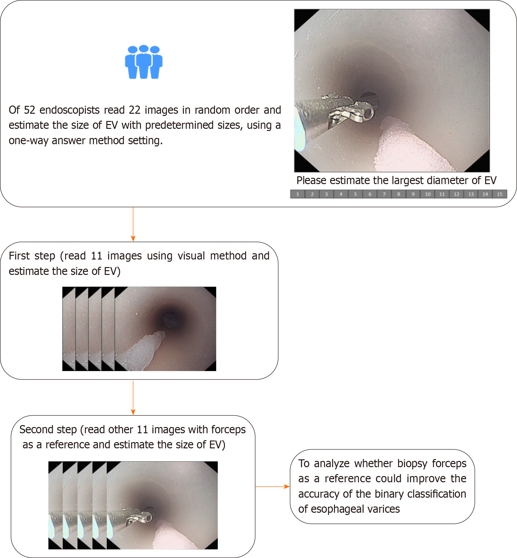
Figure 1 Flow chart of this study.
EV: Esophageal varices.
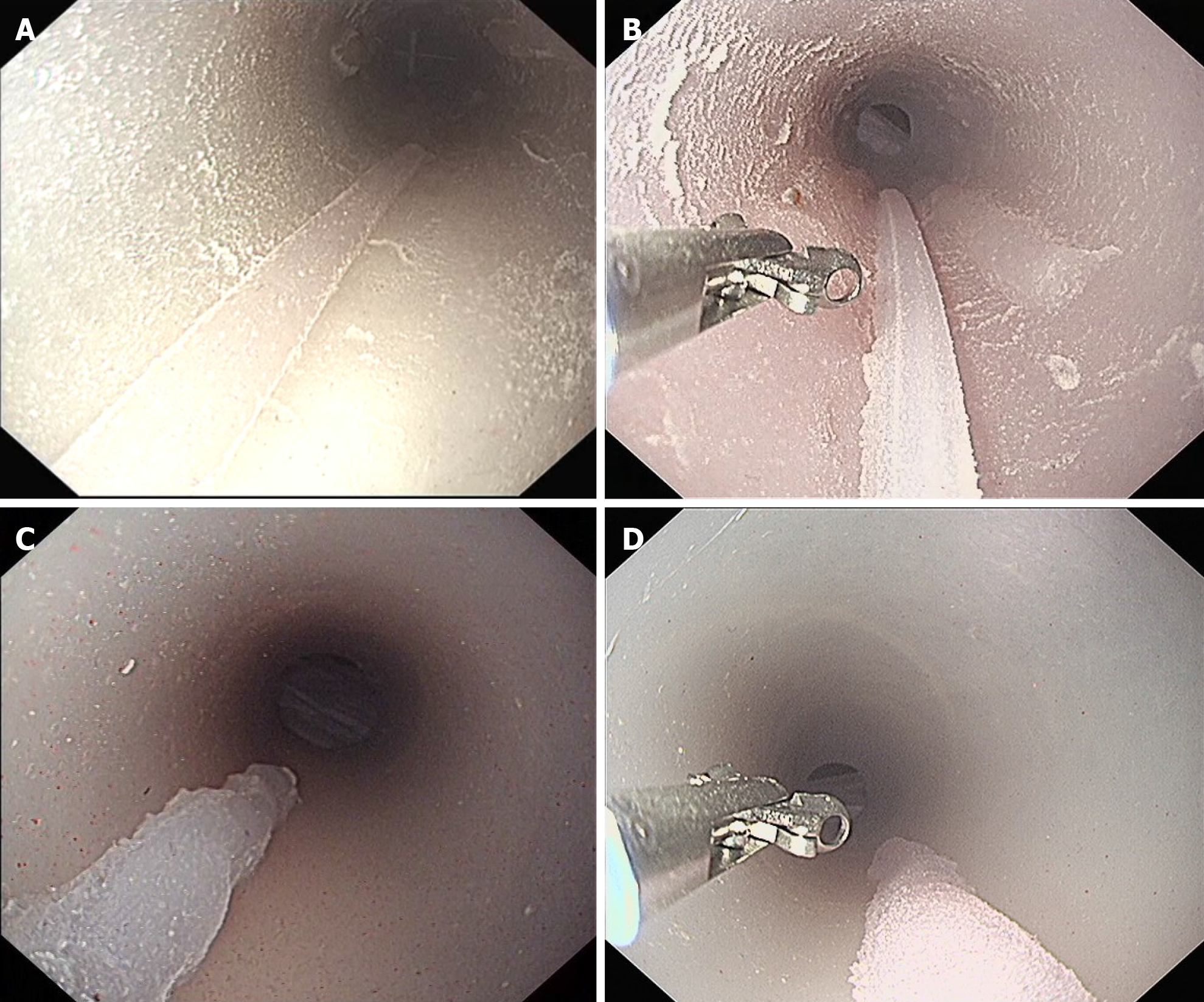
Figure 2 Examples of images for online testing in this study.
A: The actual size of the varices in picture (traditional visual method) was 3 mm; B: The actual size of the varices in the picture (biopsy forceps as a reference) was 3 mm; C: The actual size of the varices in the picture (visual method) was 7 mm; D: The actual size of the varices in the picture (biopsy forceps method) was 7 mm.
- Citation: Duan ZH, Zhou SY. Biopsy forceps are useful for measuring esophageal varices in vitro. World J Gastrointest Surg 2024; 16(2): 539-545
- URL: https://www.wjgnet.com/1948-9366/full/v16/i2/539.htm
- DOI: https://dx.doi.org/10.4240/wjgs.v16.i2.539


