Copyright
©The Author(s) 2024.
World J Gastrointest Surg. Feb 27, 2024; 16(2): 409-418
Published online Feb 27, 2024. doi: 10.4240/wjgs.v16.i2.409
Published online Feb 27, 2024. doi: 10.4240/wjgs.v16.i2.409
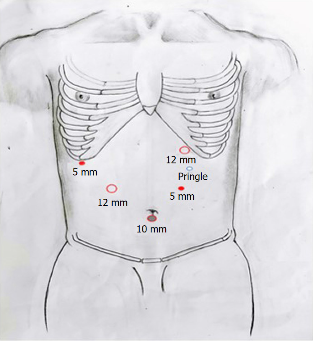
Figure 1 Trocar placement.
Two 12-mm trocars, two 5-mm trocars, and one 10-mm trocar were used. A 3-mm incision was made for intermittent Pringle's maneuver.
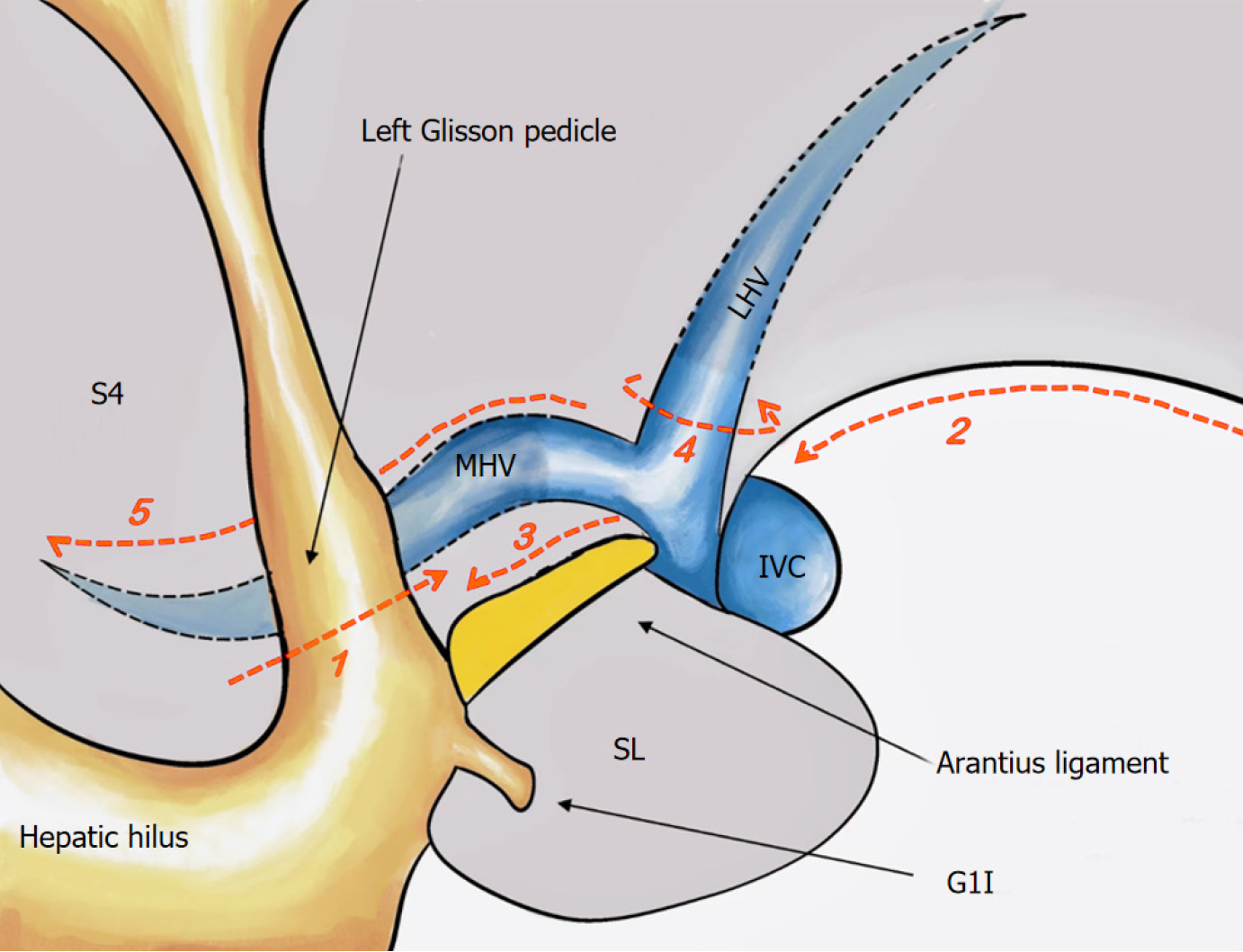
Figure 2 Standard procedure of the cranial-dorsal strategy for laparoscopic left-hemihepatectomy along the Arantius ligament.
(1) Treatment of the left Glissonean pedicle; (2) hepatic mobilization; (3) separation of the lobe through the dissection of the ligament plane; (4) identification of the middle hepatic vein (MHV) and left-hepatic vein; and (5) 180° exposure of the MHV on the hepatic section. MHV: Middle hepatic vein; IVC: Inferior vena cava; LHV: Left-hepatic vein; SL: Spiegelian lobe; S4: Segment 4 of liver.
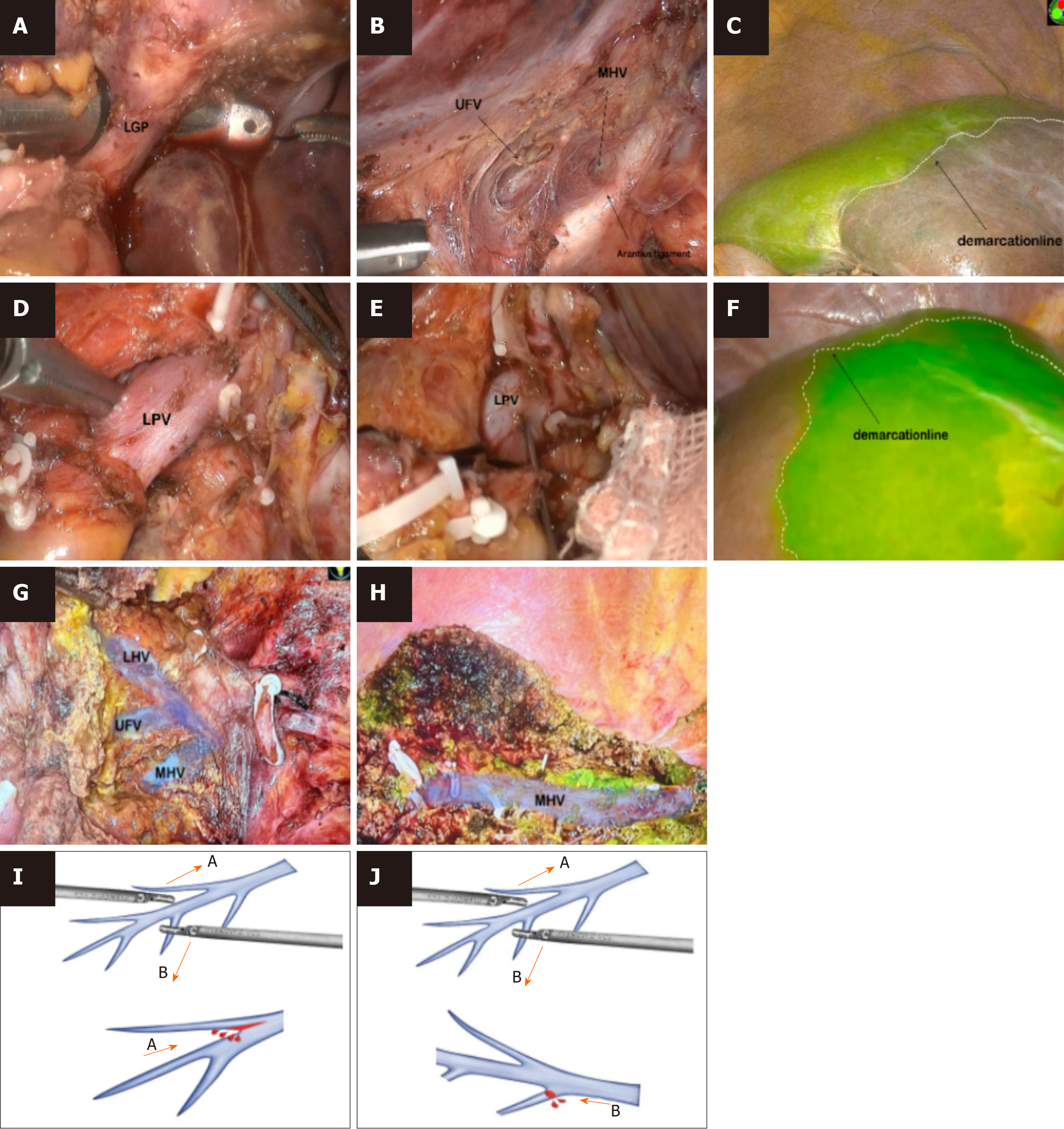
Figure 3 Intraoperative images during the first operation.
A: Extrahepatic left-Glissonean pedicle isolation; B: The middle hepatic vein (MHV) and left hepatic vein (LHV) can be well controlled and pre-treated by dissecting the caudal edge of the Arantius ligament; C: The demarcation line confirmed by indocyanine green (ICG) fluorescence; D: Intrathecal dissection of the left portal vein (LPV); E: Puncture of the LPV vein and ICG injection; F: The demarcation line confirmed by ICG fluorescence; G: Using the cranial-dorsal strategy, the LHV and MHV can be exposed from the cranial side, and the umbilical fissure vein is often lined on the dorsal side of the LHV; H: The MHV was exposed at 180° throughout the entire procedure, and there was no ICG signal on the hepatic section; I: Exposing the hepatic vein from the periphery to the MHV root would cause split injuries; J: Using the cranial-dorsal strategy, proper exposure of the MHV can reduce bleeding. ICG: Indocyanine green; LGP: Left-Glissonean pedicle; LHV: Left hepatic vein; LPV: Left portal vein; MHV: Middle hepatic vein; UFV: Umbilical fissure vein.
- Citation: Wang XR, Li XJ, Wan DD, Zhang Q, Liu TX, Shen ZW, Tong HX, Li Y, Li JW. Laparoscopic left hemihepatectomy guided by indocyanine green fluorescence: A cranial-dorsal approach. World J Gastrointest Surg 2024; 16(2): 409-418
- URL: https://www.wjgnet.com/1948-9366/full/v16/i2/409.htm
- DOI: https://dx.doi.org/10.4240/wjgs.v16.i2.409


