Copyright
©The Author(s) 2024.
World J Gastrointest Surg. Oct 27, 2024; 16(10): 3328-3333
Published online Oct 27, 2024. doi: 10.4240/wjgs.v16.i10.3328
Published online Oct 27, 2024. doi: 10.4240/wjgs.v16.i10.3328
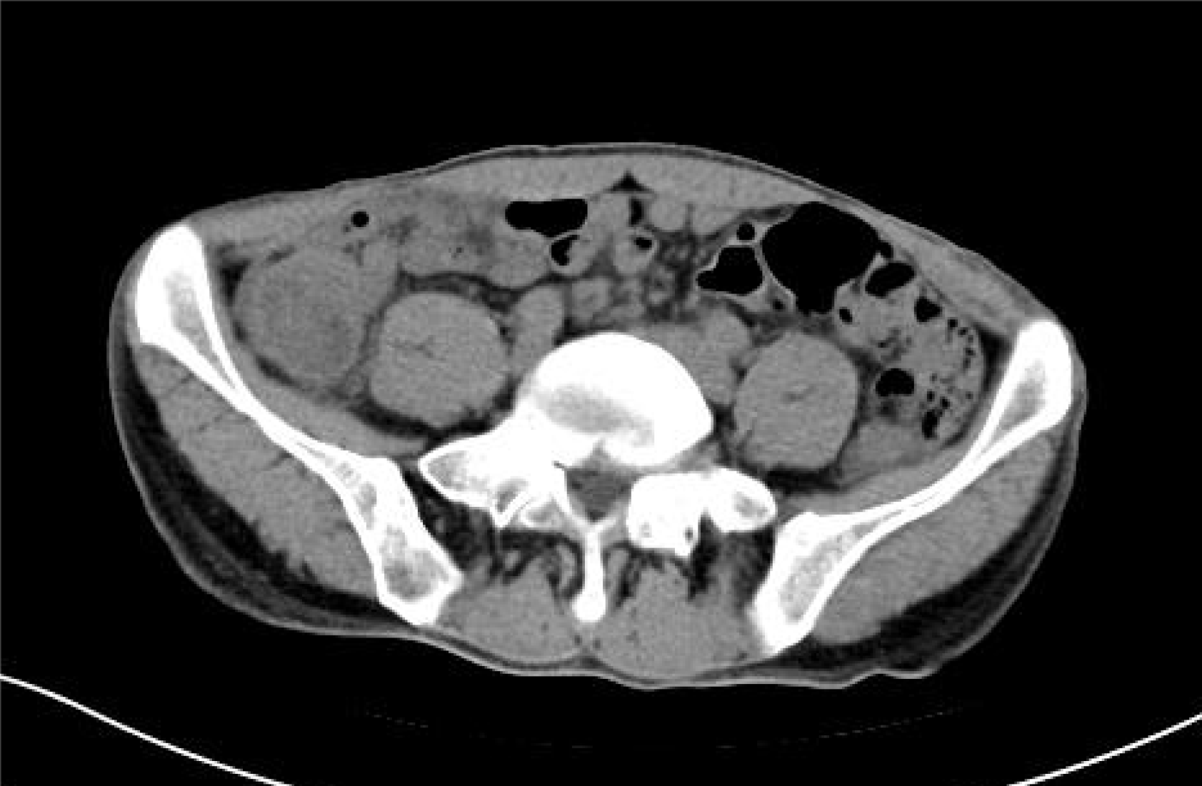
Figure 1
Preoperative abdominal computed tomography scan suggested appendicitis with localized peritonitis.
Figure 2 Intraoperative arrangement.
A: Intraoperative device placement: A 1 cm trocar (for primary operation) and a 5 mm trocar (for laparoscopic observation) were inserted into the abdominal cavity via the umbilical incision, and a needle-type grasping forceps (Approval No. Zsyjx 20140056; Hangzhou Kangji Medical Instrument Co., Ltd., Hangzhou, China) were inserted at the McBurney point; B: Surgeon’s standing position: The laparoscopic display screen was on the right side of the patient, while the surgeon stood down to the left and the assistant at the upper right; C: Intraoperative laparoscopic images: The appendix was located in the anterior position of the cecum, and the surface was hyperemic and edematous; needle-type forceps were inserted into the abdominal cavity to assist with traction of the appendix's mesentery; sterile gauze was inserted into the right iliac fossa to clear bleeding and exudation; longer Ham-o-lock clips were placed to shut the root and distal part of the appendix; and an ultrasonic scalpel was applied to cut the appendix accompanying its mesentery.
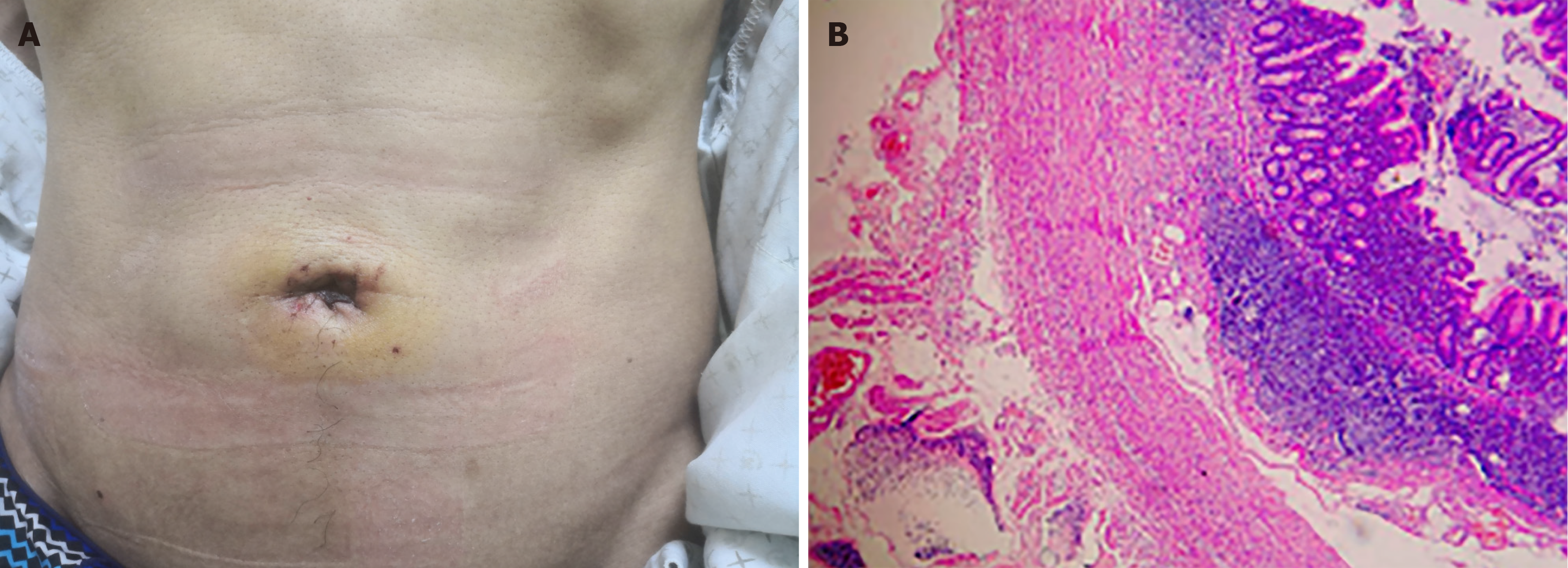
Figure 3 Postoperative outcomes.
A: Postoperative day 2 incision appearance; B: Postoperative pathologic outcome (200 × HE staining).
- Citation: Chen Y, Fan ZQ, Fu XA, Zhang XX, Yuan JQ, Guo SG. Modified technical protocol for single-port laparoscopic appendectomy using needle-type grasping forceps for acute simple appendicitis: A case report. World J Gastrointest Surg 2024; 16(10): 3328-3333
- URL: https://www.wjgnet.com/1948-9366/full/v16/i10/3328.htm
- DOI: https://dx.doi.org/10.4240/wjgs.v16.i10.3328


