Copyright
©The Author(s) 2024.
World J Gastrointest Surg. Oct 27, 2024; 16(10): 3312-3320
Published online Oct 27, 2024. doi: 10.4240/wjgs.v16.i10.3312
Published online Oct 27, 2024. doi: 10.4240/wjgs.v16.i10.3312
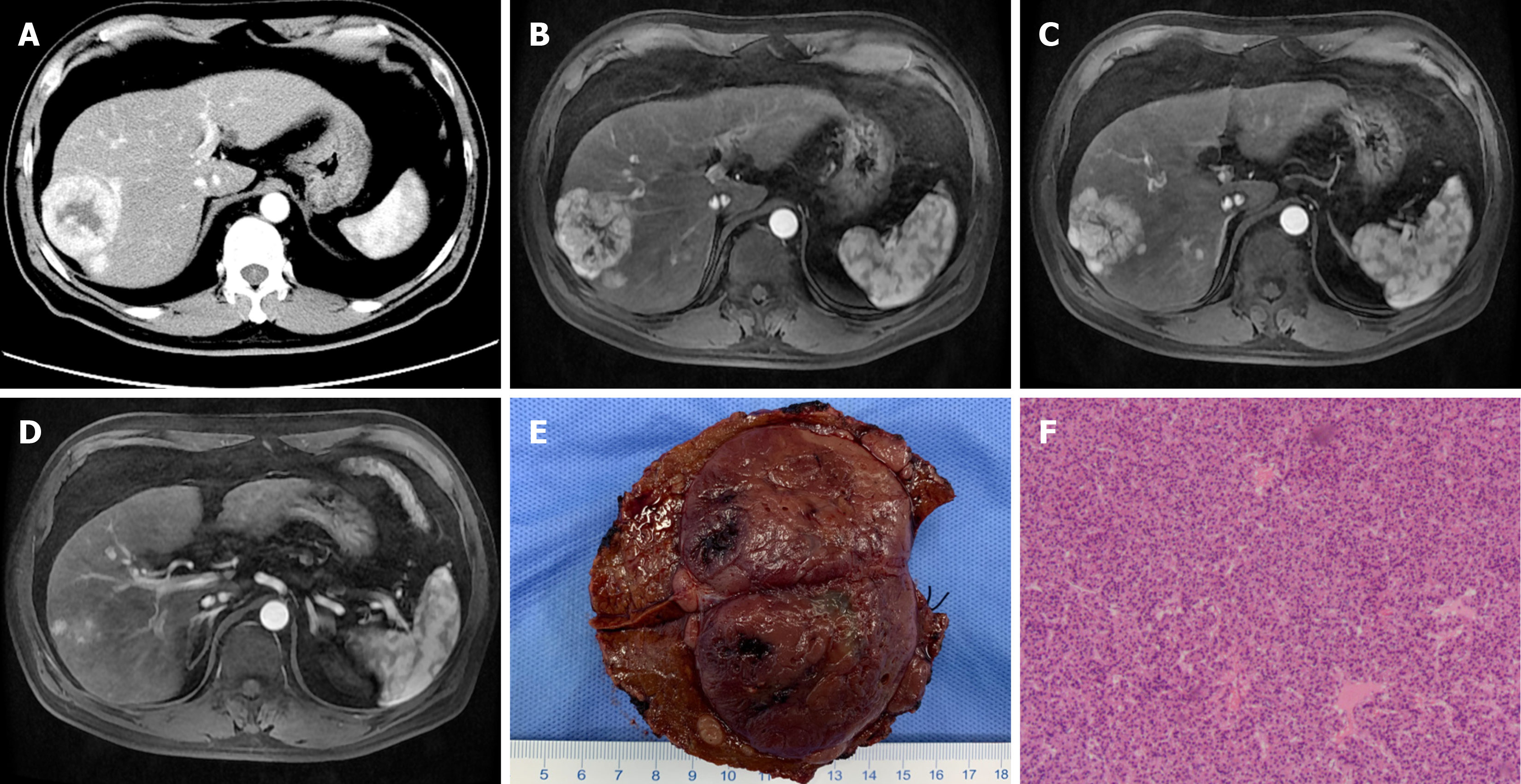
Figure 1 Preoperative computed tomography and magnetic resonance imaging of case 1.
A-D: Contrast-enhanced computed tomography and magnetic resonance imaging of the liver revealed a 65 mm × 55 mm lesion in the S7/8 lobe with multiple intrahepatic metastases in the arterial phase; E: Macroscopic findings of the tumor revealed the major lesion with satellite micronodules (with a diameter of 0.3-1 cm); F: Microscopic diagnosis revealed moderately differentiated hepatocellular carcinoma with negative surgical margins (magnification, 40 ×).
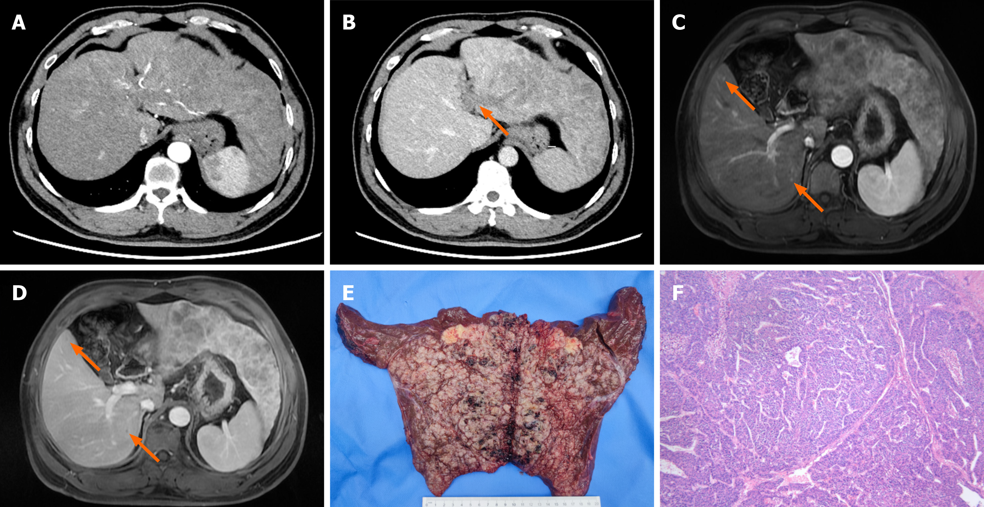
Figure 2 Preoperative computed tomography and magnetic resonance imaging of case 2.
A and B: Contrast-enhanced computed tomography showed an 18 cm × 15 cm massive lesion in the left liver, with multiple intrahepatic foci, contrast enhancement in the arterial phase and relatively low density in the delayed phase, tumor thrombus formation in the left branch of the portal vein (arrow); C and D: T1-weighted enhanced magnetic resonance imaging confirmed two of the multiple intrahepatic foci, contrast enhancement in the arterial phase (arrow), and relatively low signal in the portal phase (arrow); E: Macroscopic findings of the tumor showed the major lesion accompanied by multiple subfoci; F: Microscopic diagnosis showed moderately differentiated hepatocellular carcinoma with microvascular tumor thrombus and negative surgical margins (magnification, 40 ×).
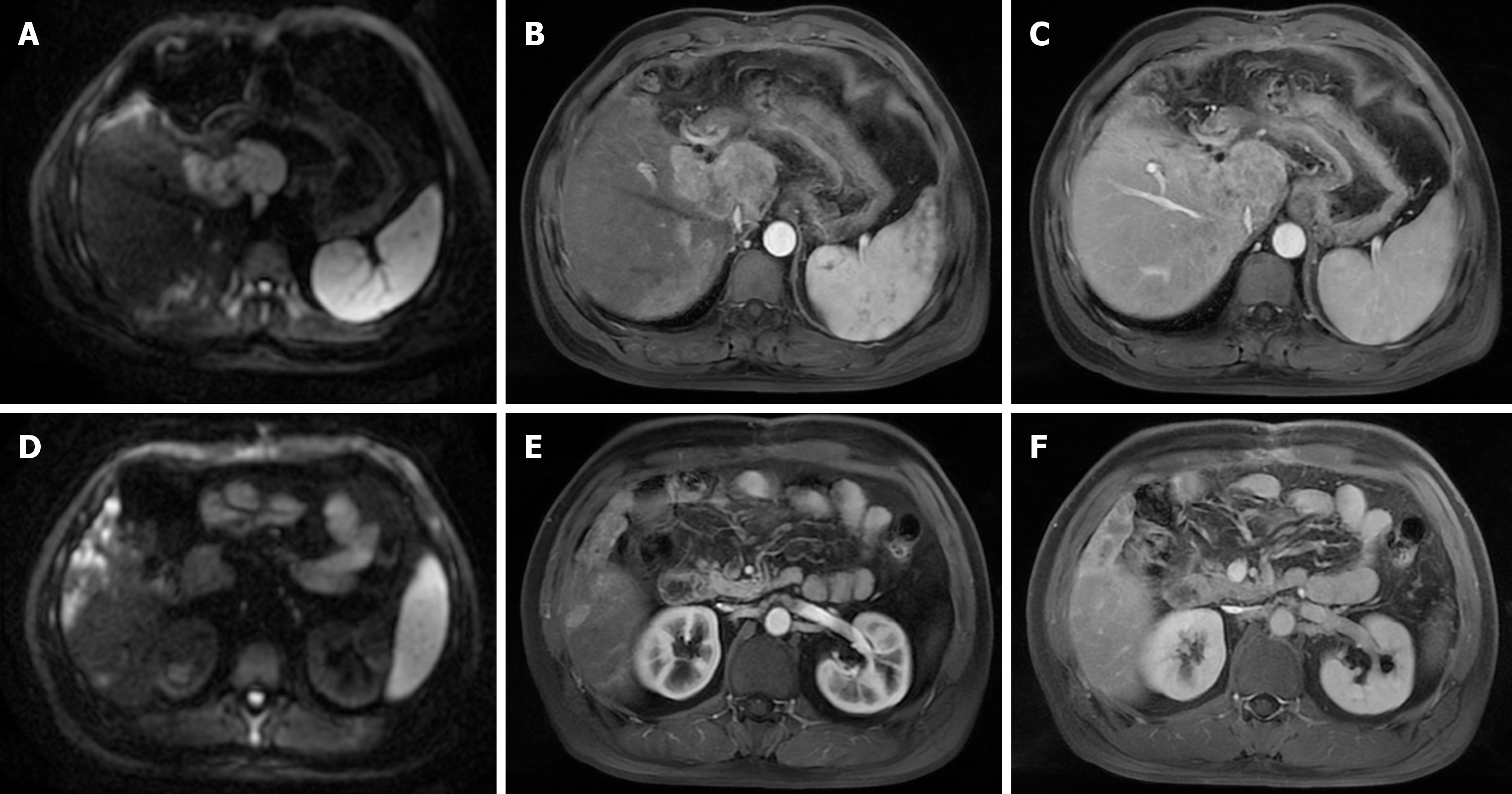
Figure 3 Post-operative magnetic resonance imaging of case 2 (1-month).
A-C: T1-weighted enhanced magnetic resonance imaging (MRI) indicated new focal hepatocellular carcinoma recurrence in the caudate lobe, which revealed a high signal in diffusion-weighted imaging, contrast enhancement in the arterial phase and relatively lower signal in the portal phase; D-F: T1-weighted enhanced MRI revealed multiple intrahepatic foci in the remaining right liver, with a high signal in diffusion-weighted imaging, contrast enhancement in the arterial phase and relatively lower signal in the hepatobiliary phase.
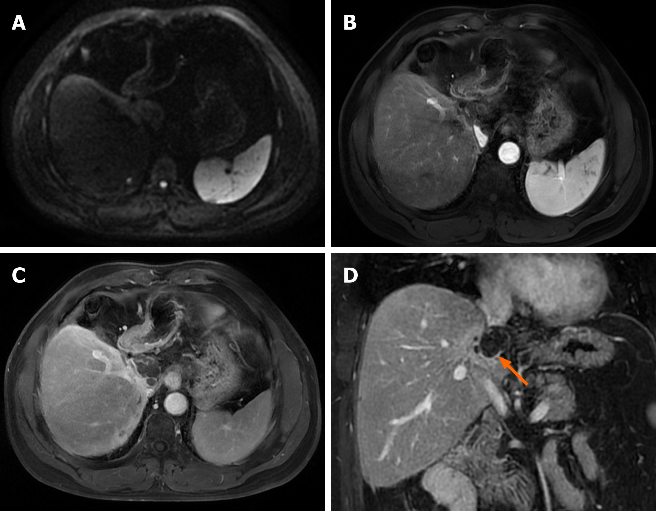
Figure 4 Post-operative magnetic resonance imaging of case 2 (6 months).
A: Diffusion-weighted phase revealed no obvious tumor activity in the residual liver; B and C: T1-weighted enhanced magnetic resonance imaging showed extensive tumor necrosis in the caudate lobe, and local slight enhancement around the tumor showing high signal in arterial phase and venous phase; D: Coronal delayed phase confirmed extensive tumor necrosis in the caudate lobe with marginal local slight enhancement (arrow) and no tumor activity in the remaining right liver.
- Citation: Zhu YB, Qin JY, Zhang TT, Zhang WJ, Ling Q. Reassessment of palliative surgery in conversion therapy of previously unresectable hepatocellular carcinoma: Two case reports and review of literature. World J Gastrointest Surg 2024; 16(10): 3312-3320
- URL: https://www.wjgnet.com/1948-9366/full/v16/i10/3312.htm
- DOI: https://dx.doi.org/10.4240/wjgs.v16.i10.3312


