Copyright
©The Author(s) 2024.
World J Gastrointest Surg. Oct 27, 2024; 16(10): 3104-3113
Published online Oct 27, 2024. doi: 10.4240/wjgs.v16.i10.3104
Published online Oct 27, 2024. doi: 10.4240/wjgs.v16.i10.3104
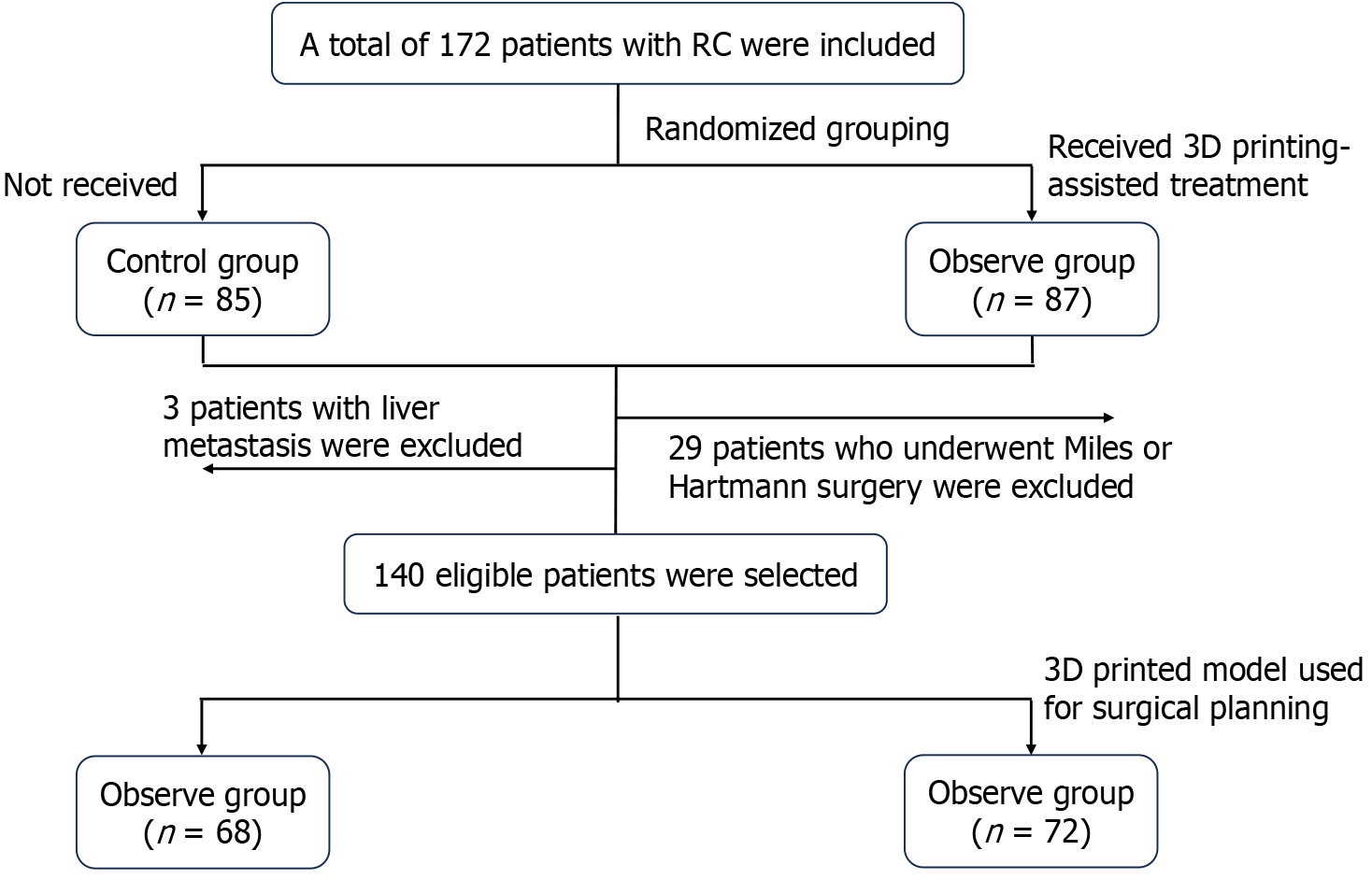
Figure 1 Flowchart of this retrospective case control study.
RC: Rectal cancer; 3D: Three dimensional.
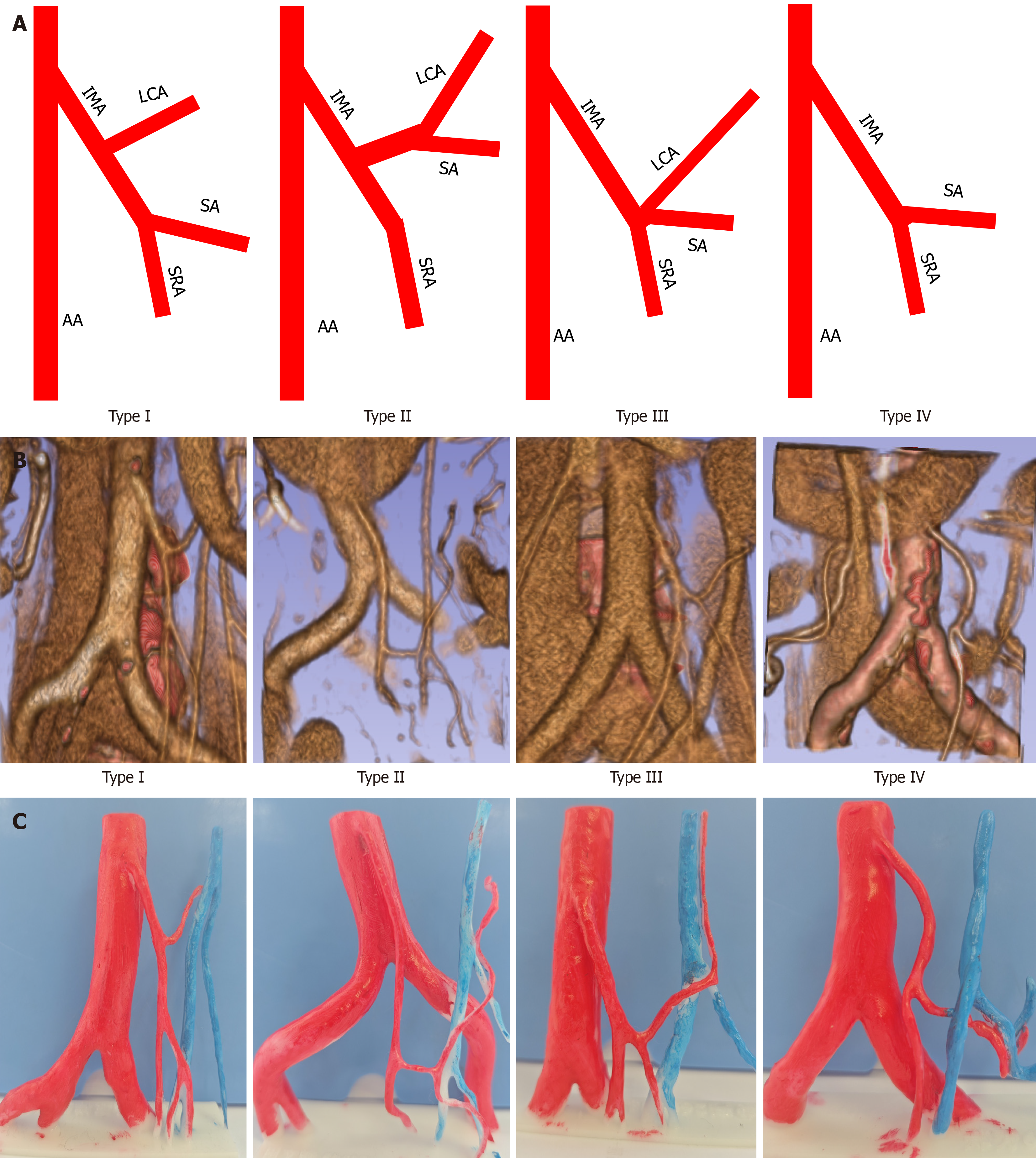
Figure 2 Overview of the four types of inferior mesenteric artery classification.
A: Schematic diagrams; B: Based on enhanced computed tomography display; C: Display using three-dimensional printing models. AA: Abdominal aorta; IMA: Inferior mesenteric artery; LCA: Left colic artery; SA: Sigmoid artery; SRA: Superior rectal artery.
Figure 3 Clinical application of three-dimensional inferior mesenteric artery model in laparoscopic radical resection with preservation of the left colic artery.
A: Comparison between the three-dimensional printed model and intraoperative photograph (inferior mesenteric artery type I); B: Comparison between the three-dimensional printed model and intraoperative photograph (inferior mesenteric artery type III). AA: Abdominal aorta; IMA: Inferior mesenteric artery; LCA: Left colic artery; IMV: Inferior mesenteric vein; SA: Sigmoid artery; SRA: Superior rectal artery.
- Citation: Zhao ZX, Hu ZJ, Yao RD, Su XY, Zhu S, Sun J, Yao Y. Three-dimensional printing for preoperative rehearsal and intraoperative navigation during laparoscopic rectal cancer surgery with left colic artery preservation. World J Gastrointest Surg 2024; 16(10): 3104-3113
- URL: https://www.wjgnet.com/1948-9366/full/v16/i10/3104.htm
- DOI: https://dx.doi.org/10.4240/wjgs.v16.i10.3104


