Copyright
©The Author(s) 2024.
World J Gastrointest Surg. Jan 27, 2024; 16(1): 21-28
Published online Jan 27, 2024. doi: 10.4240/wjgs.v16.i1.21
Published online Jan 27, 2024. doi: 10.4240/wjgs.v16.i1.21
Figure 1 A normal appendix.
A: On the longitudinal view, the appendix appears as a blind-ending structure extending from the cecum in the abdominal cavity, the mural structures are clearly visible, there is no filling in the lumen, and the mucosa appears slightly hyperechoic with a thread-like appearance; B: On the transverse view, the appendix appears as an ovoid shape, the mural structures are visible, there is no filling in the lumen, and the ratio of the cross diameters on the transverse section is 5.9/5.3 = 1.11. Subsequent ultrasound examination revealed that the patient had a right ureter stone (not shown).
Figure 2 Acute appendicitis.
The images obtained using ultrasound from a 12-year-old female patient who presented with right inferior quadrant abdominal pain, tenderness of the abdominal wall, and abdominal guarding. A: On the longitudinal view, the appendix appears as a bent, blind-ending tubular structure extending from the cecum, the mural structures of the appendix are unclear, and there are heterogeneous complex echogenic fillings in the lumen; B: On the transverse view, the appendix appears as a round shape, with dilating lumen, there are heterogeneous complex echogenic fillings in the lumen, and the ratio of the cross diameters on the transverse section is 10/9.8 = 1.02. Postoperative histopathology confirmed that the patient had acute phlegmonous appendicitis.
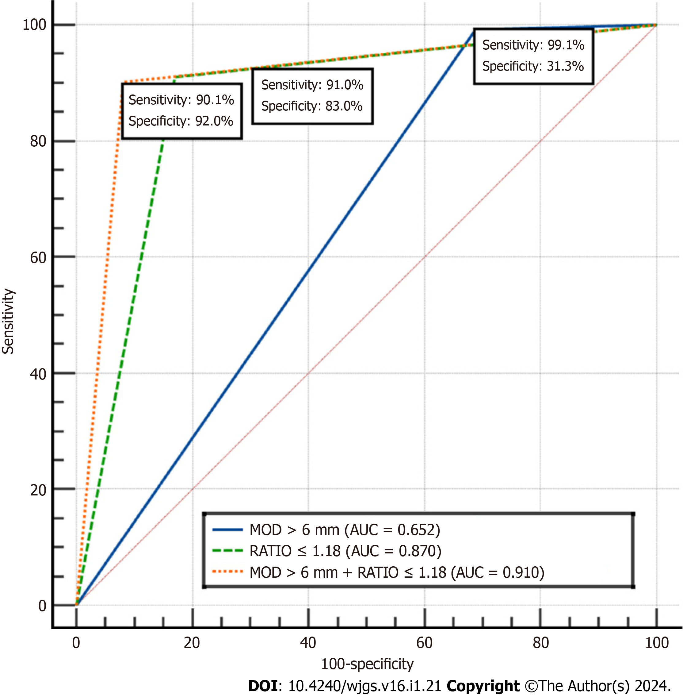
Figure 3 The receiver operating characteristic curves of ratio of the cross diameters on transverse section of the appendix, maximum outer diameter> 6 mm, and maximum outer diameter > 6 mm + ratio of the cross diameters on transverse section of the appendix.
MOD: Maximum outer diameter; AUC: Area under the receiver operating characteristic curve; RATIO: Ratio of the cross diameters of the appendix.
- Citation: Gu FW, Wu SZ. Added value of ratio of cross diameters of the appendix in ultrasound diagnosis of acute appendicitis. World J Gastrointest Surg 2024; 16(1): 21-28
- URL: https://www.wjgnet.com/1948-9366/full/v16/i1/21.htm
- DOI: https://dx.doi.org/10.4240/wjgs.v16.i1.21


