Copyright
©The Author(s) 2023.
World J Gastrointest Surg. Sep 27, 2023; 15(9): 2083-2088
Published online Sep 27, 2023. doi: 10.4240/wjgs.v15.i9.2083
Published online Sep 27, 2023. doi: 10.4240/wjgs.v15.i9.2083
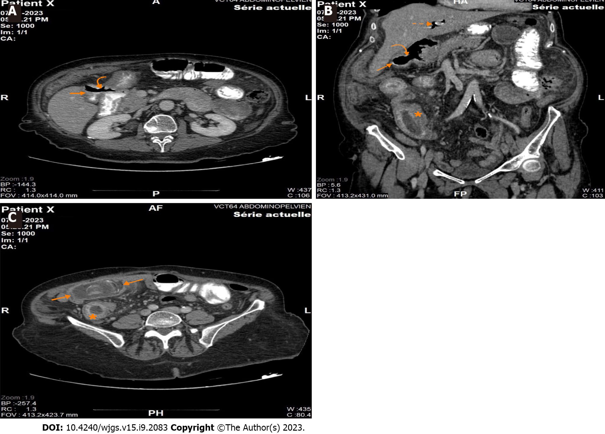
Figure 1 Computed tomography scan.
A: Axial section of contrast-enhanced computed tomography (CT) shows a gallbladder containing a hydro-aeric level with passage of ingested contrast medium (straight arrow) associated to a visible communication of 15 mm between the gallbladder and the duodenal bulb (curved arrow); B: Coronal reconstructed image (using multiplanar reconstruction) of contrast-enhanced CT shows the defect between the gallbladder and the duodenal bulb (curved arrow), the gallbladder filled with air (straight arrow) as well as pneumobilia within the intrahepatic biliary ducts (dotted straight arrow). Note that the distal ileum is mildly dilated, markedly thickened with increased mucosal enhancement (asterisks); C: Axial section of contrast-enhanced CT shows a gallstone measuring almost 45 mm (two arrows) impacted in the distal ileum, with marked thickening and increased mucosal enhancement within the upstream ileum (asterisks).
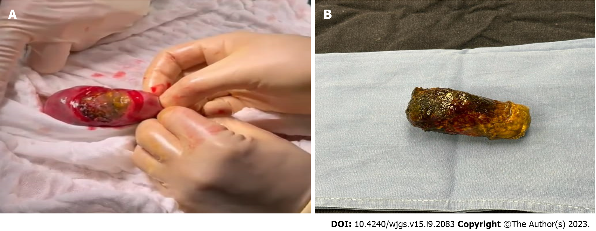
Figure 2 Gallstone.
A: Intraoperative photo of gallstone after enterolithotomy; B: Gallstone.
- Citation: El Feghali E, Akel R, Chamaa B, Kazan D, Chakhtoura G. Surgical management of gallstone ileus after one anastomosis gastric bypass: A case report. World J Gastrointest Surg 2023; 15(9): 2083-2088
- URL: https://www.wjgnet.com/1948-9366/full/v15/i9/2083.htm
- DOI: https://dx.doi.org/10.4240/wjgs.v15.i9.2083


