Copyright
©The Author(s) 2023.
World J Gastrointest Surg. Sep 27, 2023; 15(9): 2074-2082
Published online Sep 27, 2023. doi: 10.4240/wjgs.v15.i9.2074
Published online Sep 27, 2023. doi: 10.4240/wjgs.v15.i9.2074
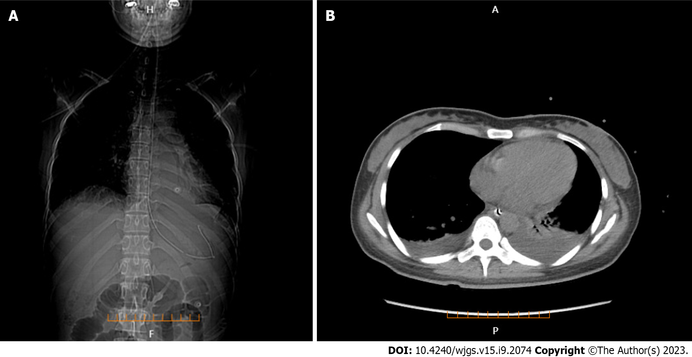
Figure 1 Lung computed tomography.
A: Bilateral pleural effusion and the position of gastrointestinal decompression tube (coronal plane); B: Bilateral pleural effusion (axial plane).
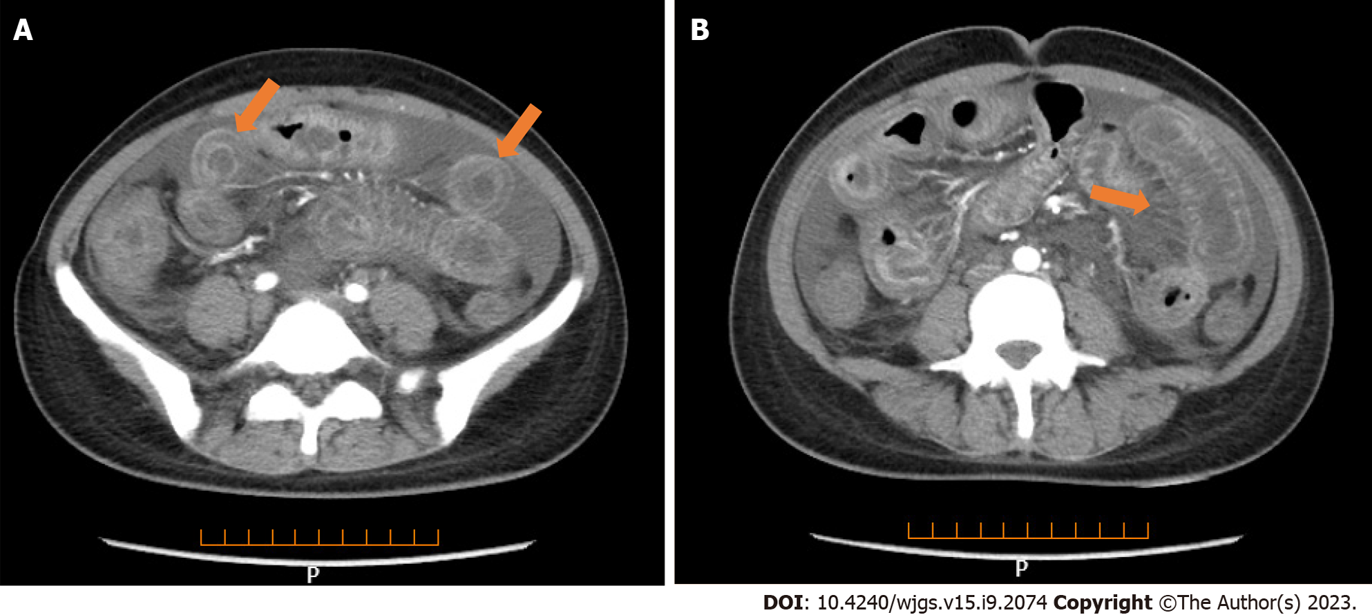
Figure 2 Full enhanced abdominal computed tomography.
A: Thickened bowel loops (target sign); B: Engorgement and increased visibility of the mesenteric vessels (comb sign).
Figure 3 Ultrasonography of the transverse colon showing thickened segmental bowel walls of approximately 5 mm.
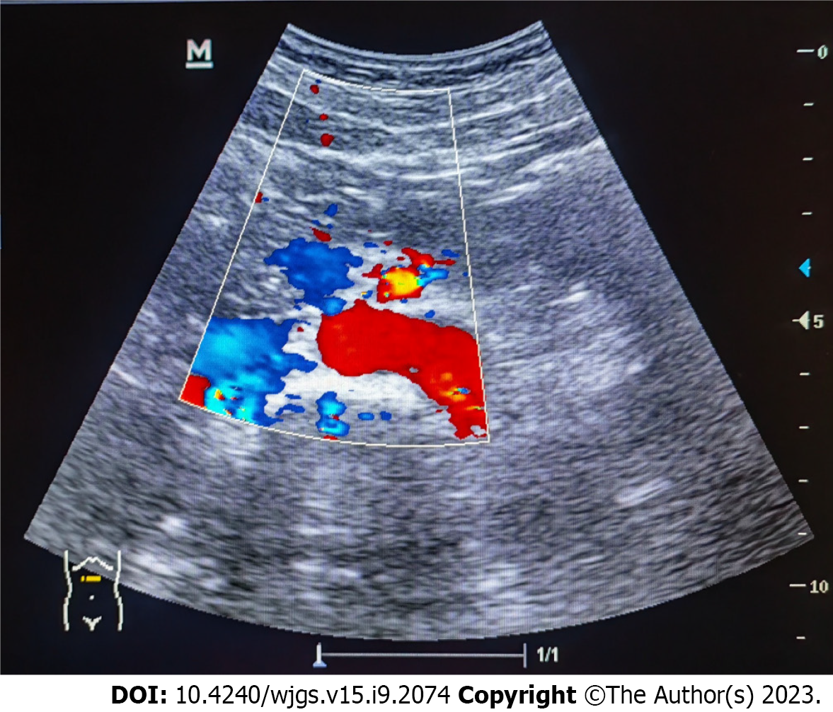
Figure 4 Ultrasonography of the superior mesenteric artery showing the main color flow of the superior mesenteric artery was well-filled without embolism.
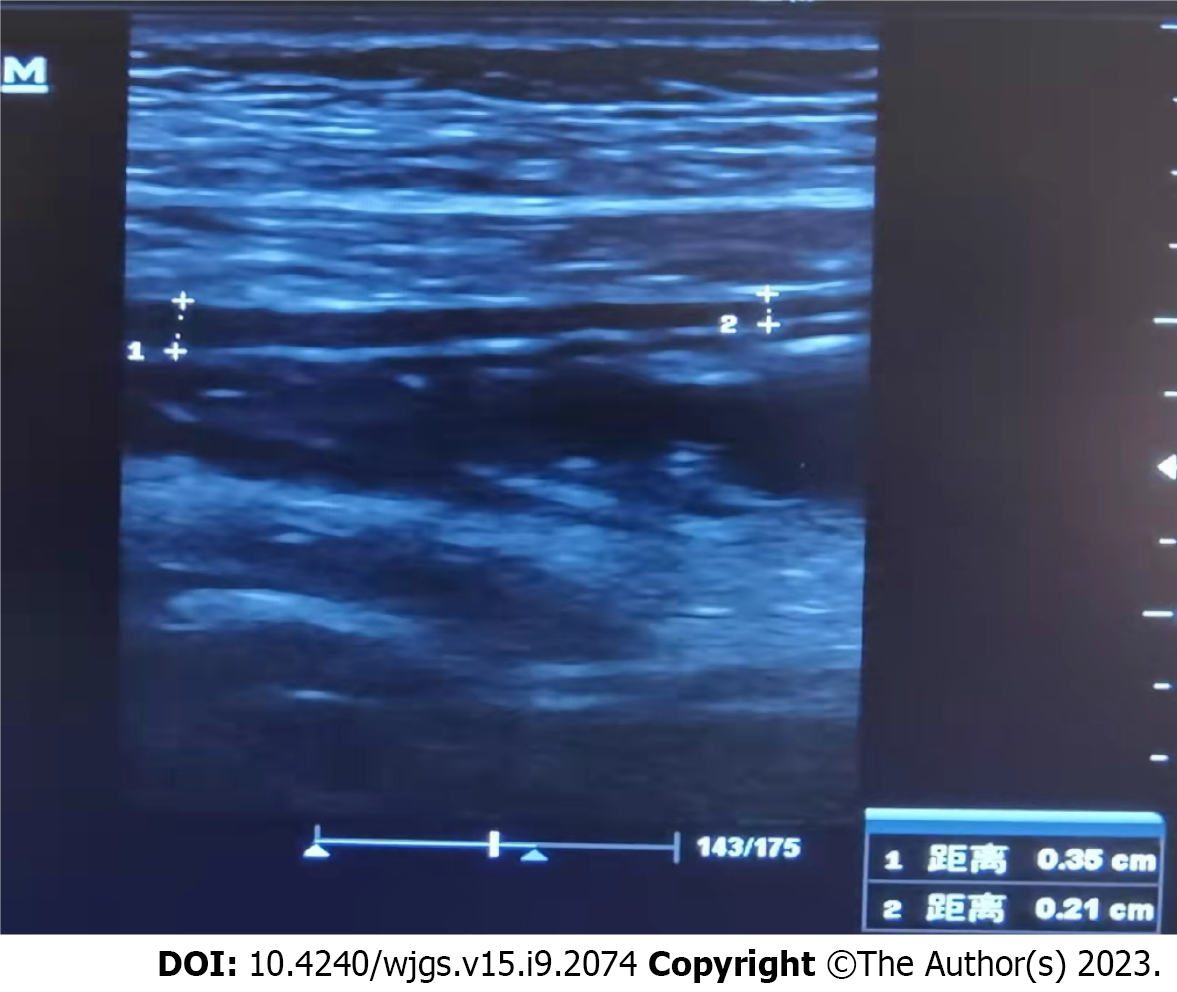
Figure 5 Ultrasonography showing the local intestinal wall with approximately 0.
35 cm at its thickest part.
- Citation: Huang H, Li P, Zhang D, Zhang MX, Yu K. Acute flare of systemic lupus erythematosus with extensive gastrointestinal involvement: A case report and review of literature. World J Gastrointest Surg 2023; 15(9): 2074-2082
- URL: https://www.wjgnet.com/1948-9366/full/v15/i9/2074.htm
- DOI: https://dx.doi.org/10.4240/wjgs.v15.i9.2074


