Copyright
©The Author(s) 2023.
World J Gastrointest Surg. Sep 27, 2023; 15(9): 2063-2073
Published online Sep 27, 2023. doi: 10.4240/wjgs.v15.i9.2063
Published online Sep 27, 2023. doi: 10.4240/wjgs.v15.i9.2063
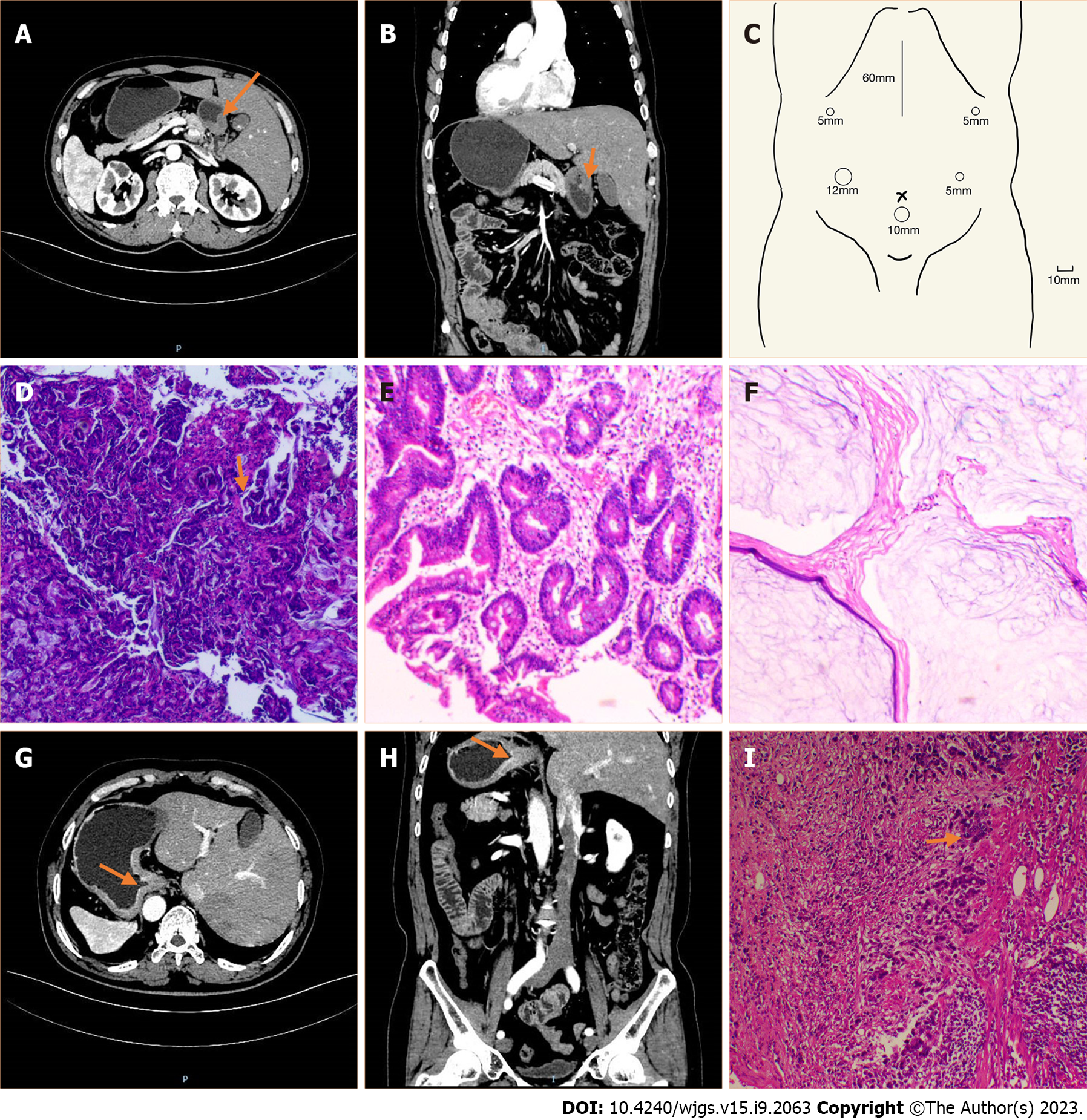
Figure 1 Imaging and pathological features of the two patients.
A–F: Case 1; A: Computed tomography (CT) image showing the inverse positioning of intra-abdominal organs; B: CT image showing antral wall thickening; C: Sites of trocar placement. A 12-mm trocar was placed in the right hypochondriac region, and the other three 5-mm trocars were placed (one each) in the right subcostal region, the right lateral abdominal region, and the left lateral abdominal region; D: Preoperative (after neoadjuvant chemotherapy) pathologic biopsies showing that tumor nests still existed; E: Postoperative pathologic biopsies showing that there were no nidi; F: Postoperative pathologic biopsies showing no nidi; G–I: Case 2; G: CT image showing the inverse positioning of all intra-abdominal organs; H: CT image revealing the thickening of the cardia and smaller curvature of gastric tissue and enlarged peri-gastric small lymph nodes; I: Postoperative pathologic biopsies showing that nidi still existed.
Figure 2 Images of surgery.
A: The surgical field showing that the spleen in the right side of the patient; B: The surgical field showing that the gallbladder and right lobe of the liver in the left side of the patient; C: The key procedure during surgery: Dissecting station 6 lymph nodes.
- Citation: Liu HB, Cai XP, Lu Z, Xiong B, Peng CW. Laparoscopy-assisted gastrectomy for advanced gastric cancer patients with situs inversus totalis: Two case reports and review of literature. World J Gastrointest Surg 2023; 15(9): 2063-2073
- URL: https://www.wjgnet.com/1948-9366/full/v15/i9/2063.htm
- DOI: https://dx.doi.org/10.4240/wjgs.v15.i9.2063


